Biomedical Engineering Department, Washington University of Saint Louis MO, Saint Louis, MO, 63130, USA.
Sci Rep. 2017 May 31;7(1):2560. doi: 10.1038/s41598-017-02458-w.
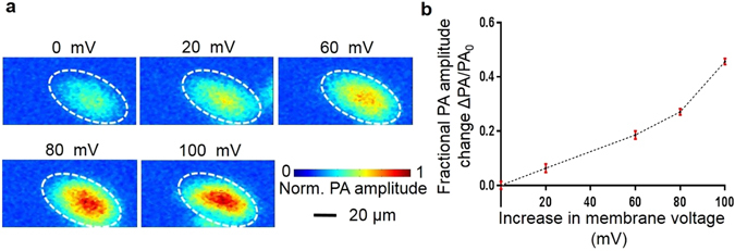
Non-invasive optical imaging of neuronal voltage response signals in live brains is constrained in depth by the optical diffusion limit, which is due primarily to optical scattering by brain tissues. Although photoacoustic tomography breaks this limit by exciting the targets with diffused photons and detecting the resulting acoustic responses, it has not been demonstrated as a modality for imaging voltage responses. In this communication, we report the first demonstration of photoacoustic voltage response imaging in both in vitro HEK-293 cell cultures and in vivo mouse brain surfaces. Using spectroscopic photoacoustic tomography at isosbestic wavelengths, we can separate voltage response signals and hemodynamic signals on live brain surfaces. By imaging HEK-293 cell clusters through 4.5 mm thick ex vivo rat brain tissue, we demonstrate photoacoustic tomography of cell membrane voltage responses beyond the optical diffusion limit. Although the current voltage dye does not immediately allow in vivo deep brain voltage response imaging, we believe our method opens up a feasible technical path for deep brain studies in the future.
在活体大脑中对神经元电压响应信号进行非侵入式光学成像是受到深度限制的,这主要是由于脑组织的光散射造成的。尽管光声断层扫描通过扩散光子激发目标并检测产生的声响应来打破这一限制,但它尚未被证明是一种用于成像电压响应的模式。在本通讯中,我们首次报道了在体外 HEK-293 细胞培养物和体内小鼠脑表面进行光声电压响应成像的结果。我们使用等消色光声断层扫描在活体脑表面分离电压响应信号和血流动力学信号。通过对 4.5mm 厚的离体大鼠脑组织中的 HEK-293 细胞簇进行成像,我们证明了细胞膜电压响应的光声断层扫描超越了光学扩散极限。虽然目前的电压染料不能立即实现活体深部脑电压响应成像,但我们相信我们的方法为未来的深部脑研究开辟了可行的技术途径。