Takano Shinichi, Fukasawa Mitsuharu, Kadokura Makoto, Shindo Hiroko, Takahashi Ei, Hirose Sumio, Fukasawa Yoshimitsu, Kawakami Satoshi, Sato Tadashi, Enomoto Nobuyuki
Shinichi Takano, Mitsuharu Fukasawa, Makoto Kadokura, Hiroko Shindo, Ei Takahashi, Sumio Hirose, Yoshimitsu Fukasawa, Satoshi Kawakami, Tadashi Sato, Nobuyuki Enomoto, First Department of Internal Medicine, Faculty of Medicine, University of Yamanashi, Yamanashi 409-3898, Japan.
World J Gastroenterol. 2017 May 14;23(18):3295-3300. doi: 10.3748/wjg.v23.i18.3295.
To assess the role of ultrasonography of submandibular glands (SGs) in the diagnosis of type 1 autoimmune pancreatitis (AIP).
Thirty-seven patients who were definitively diagnosed with type 1 AIP according to the international consensus diagnostic criteria (ICDC) for AIP at our institution between December 1990 and April 2016 were retrospectively reviewed. Findings by physical examination, ultrasonography, and scintigraphy of SGs were analyzed to reach a diagnosis based on the ICDC for AIP. The efficacy of corticosteroid treatment in the resolution of hypoechoic lesions in SGs was also evaluated by assessment with ultrasonography before and after treatment in 18 cases.
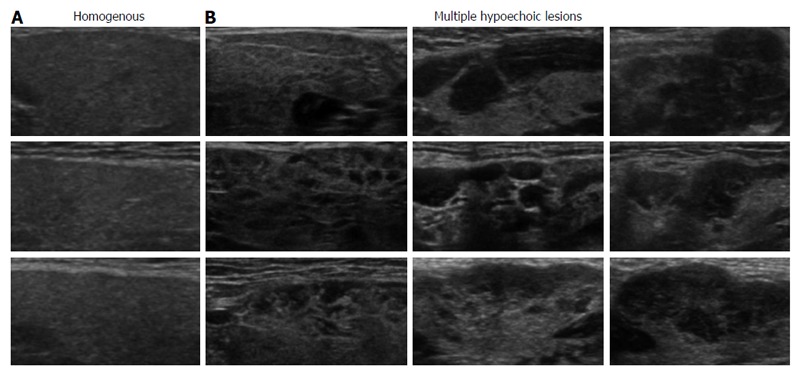
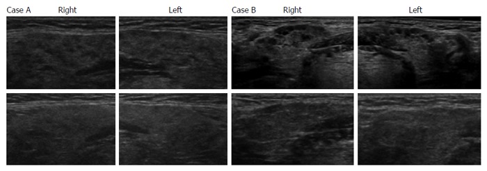
The sensitivity of multiple hypoechoic lesions in SGs by ultrasonography for the diagnosis of sialadenitis in type 1 AIP (84%) was higher than that of physical examination (46%), scintigraphy (28%), and SGs thickness (49%). Ultrasonographic evidence of hypoechoic lesions in SGs improved the definitive diagnosis of sialadenitis and type 1 AIP by the ICDC criteria in 11 (30%) and 2 (5.4%) cases, respectively. Multiple hypoechoic lesions in SGs were resolved or disappear by corticosteroid administration in 14 of 16 cases with hypoechoic lesions in SGs, whereas the ultrasonographic findings in the remaining 2 cases with hypoechoic lesions in SGs and the 2 cases with homogenous SG parenchyma remained unchanged after corticosteroid administration.
SG ultrasonography to detect multiple hypoechoic lesions might be useful for type 1 AIP diagnosis by improving diagnostic accuracy together with the ICDC sialadenitis criteria.
评估下颌下腺超声检查在1型自身免疫性胰腺炎(AIP)诊断中的作用。
回顾性分析1990年12月至2016年4月在我院根据AIP国际共识诊断标准(ICDC)确诊为1型AIP的37例患者。分析体格检查、超声检查和下颌下腺闪烁扫描的结果,以根据ICDC诊断AIP。还通过对18例患者治疗前后的超声评估,评价皮质类固醇治疗对下颌下腺低回声病变消退的疗效。
超声检查发现下颌下腺多发低回声病变对1型AIP涎腺炎诊断的敏感性(84%)高于体格检查(46%)、闪烁扫描(28%)和下颌下腺厚度(49%)。下颌下腺低回声病变的超声证据分别在11例(30%)和2例(5.4%)中提高了根据ICDC标准对涎腺炎和1型AIP的明确诊断。16例下颌下腺有低回声病变的患者中,14例经皮质类固醇治疗后低回声病变消退或消失,而其余2例下颌下腺有低回声病变的患者和2例下颌下腺实质均匀的患者在皮质类固醇治疗后超声表现未改变。
通过检测多发低回声病变的下颌下腺超声检查,结合ICDC涎腺炎标准提高诊断准确性,可能有助于1型AIP的诊断。