Pinto Sara, Cunha Carolina, Barbosa Marta, Vaz Ana R, Brites Dora
Neuron Glia Biology in Health and Disease, Research Institute for Medicines (iMed.ULisboa), Faculty of Pharmacy, Universidade de LisboaLisbon, Portugal.
Department of Biochemistry and Human Biology, Faculty of Pharmacy, Universidade de LisboaLisbon, Portugal.
Front Neurosci. 2017 May 17;11:273. doi: 10.3389/fnins.2017.00273. eCollection 2017.
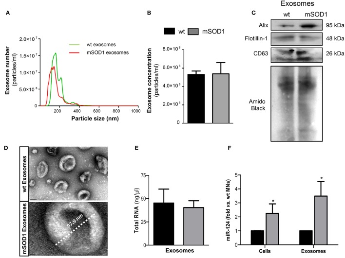
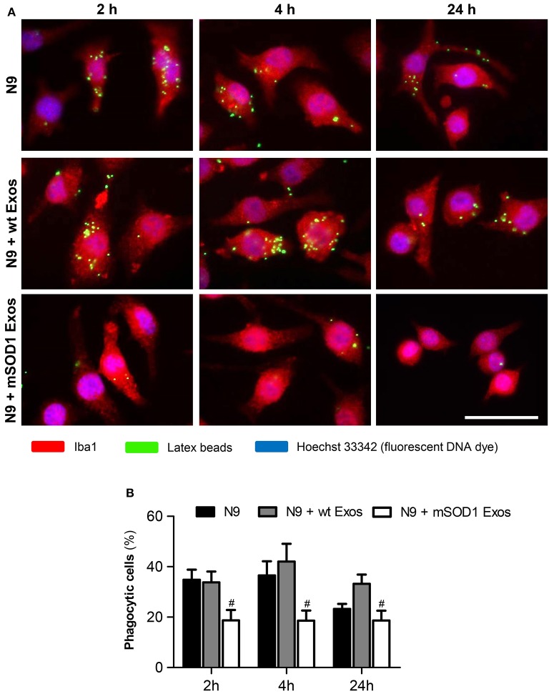
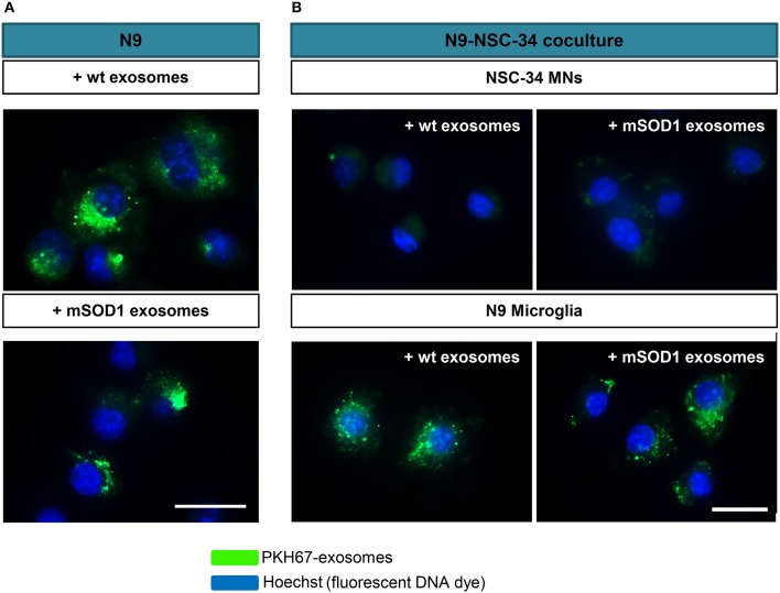
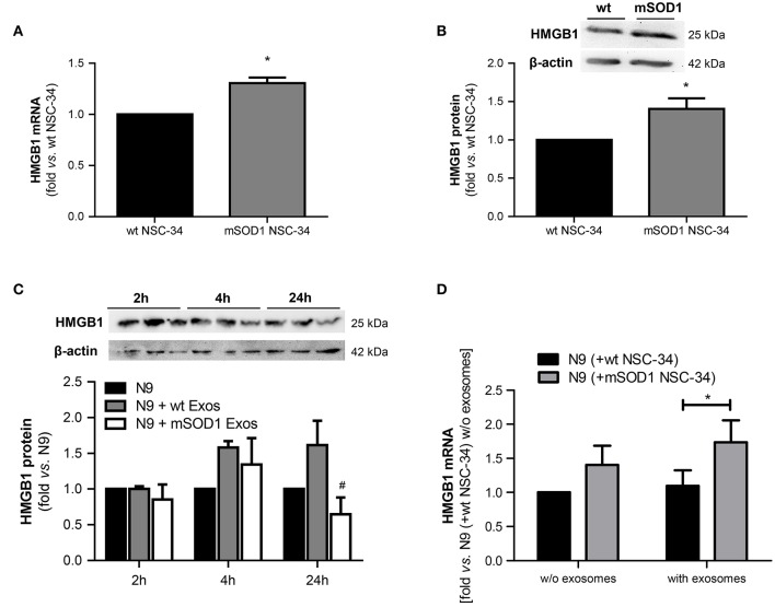
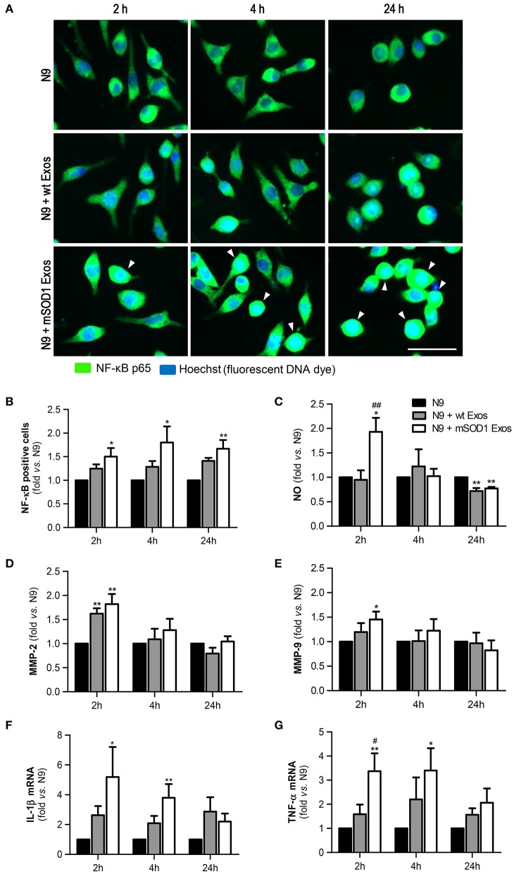
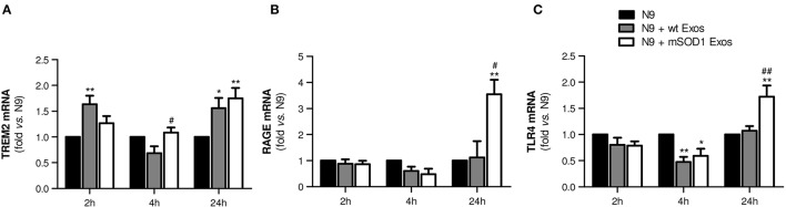
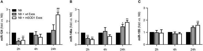
Amyotrophic lateral sclerosis (ALS) is a fatal adult-onset neurodegenerative disorder affecting motor neurons (MNs). Evidences indicate that ALS is a non-cell autonomous disease in which glial cells participate in both disease onset and progression. Exosomal transfer of mutant copper-zinc superoxide dismutase 1 (mSOD1) from cell-to-cell was suggested to contribute to disease dissemination. Data from our group and others showed that exosomes from activated cells contain inflammatory-related microRNAs (inflamma-miRNAs) that recapitulate the donor cell. While glia-derived exosomes and their effects in neurons have been addressed by several studies, only a few investigated the influence of motor neuron (MN)-derived exosomes in other cell function, the aim of the present study. We assessed a set of inflamma-miRs in NSC-34 MN-like cells transfected with mutant SOD1(G93A) and extended the study into their derived exosomes (mSOD1 exosomes). Then, the effects produced by mSOD1 exosomes in the activation and polarization of the recipient N9 microglial cells were investigated. Exosomes in coculture with N9 microglia and NSC-34 cells [either transfected with either wild-type (wt) human SOD1 or mutant SOD1(G93A)] showed to be transferred into N9 cells. Increased miR-124 expression was found in mSOD1 NSC-34 cells and in their derived exosomes. Incubation of mSOD1 exosomes with N9 cells determined a sustained 50% reduction in the cell phagocytic ability. It also caused a persistent NF-kB activation and an acute generation of NO, MMP-2, and MMP-9 activation, as well as upregulation of IL-1β, TNF-α, MHC-II, and iNOS gene expression, suggestive of induced M1 polarization. Marked elevation of IL-10, Arginase 1, TREM2, RAGE, and TLR4 mRNA levels, together with increased miR-124, miR-146a, and miR-155, at 24 h incubation, suggest the switch to mixed M1 and M2 subpopulations in the exosome-treated N9 microglial cells. Exosomes from mSOD1 NSC-34 MNs also enhanced the number of senescent-like positive N9 cells. Data suggest that miR-124 is translocated from the mSOD1 MNs to exosomes, which determine early and late phenotypic alterations in the recipient N9-microglial cells. In conclusion, modulation of the inflammatory-associated miR-124, in mSOD1 NSC-34 MNs, with potential benefits in the cargo of their exosomes may reveal a promising therapeutic strategy in halting microglia activation and associated effects in MN degeneration.
肌萎缩侧索硬化症(ALS)是一种影响运动神经元(MNs)的致命性成人起病的神经退行性疾病。有证据表明,ALS是一种非细胞自主性疾病,其中胶质细胞参与疾病的发生和进展。突变型铜锌超氧化物歧化酶1(mSOD1)在细胞间的外泌体转移被认为有助于疾病传播。我们团队和其他团队的数据表明,活化细胞来源的外泌体含有可重现供体细胞的炎症相关微小RNA(炎症微小RNA)。虽然已有多项研究探讨了胶质细胞来源的外泌体及其对神经元的影响,但只有少数研究调查了运动神经元(MN)来源的外泌体对其他细胞功能的影响,这也是本研究的目的。我们评估了用突变型SOD1(G93A)转染的NSC-34 MN样细胞中的一组炎症微小RNA,并将研究扩展到它们衍生的外泌体(mSOD1外泌体)。然后,研究了mSOD1外泌体对受体N9小胶质细胞活化和极化的影响。与N9小胶质细胞和NSC-34细胞(分别用野生型(wt)人SOD1或突变型SOD1(G93A)转染)共培养的外泌体显示被转移到N9细胞中。在mSOD1 NSC-34细胞及其衍生的外泌体中发现miR-124表达增加。用mSOD1外泌体孵育N9细胞导致细胞吞噬能力持续降低50%。它还导致持续的NF-κB激活以及NO、MMP-2和MMP-9的急性产生,以及IL-1β、TNF-α、MHC-II和iNOS基因表达上调,提示诱导M1极化。在孵育24小时时,IL-10、精氨酸酶1、TREM2、RAGE和TLR4 mRNA水平显著升高,同时miR-124、miR-146a和miR-155增加,表明外泌体处理的N9小胶质细胞转变为混合的M1和M2亚群。来自mSOD1 NSC-34 MNs的外泌体也增加了衰老样阳性N9细胞的数量。数据表明,miR-124从mSOD1 MNs转运到外泌体中,这决定了受体N9小胶质细胞的早期和晚期表型改变。总之,调节mSOD1 NSC-34 MNs中与炎症相关的miR-124,可能对其外泌体的货物有潜在益处,这可能揭示一种有前景的治疗策略,以阻止小胶质细胞活化及MN变性中的相关影响。