Kiraly Alex J, Roberts Andrea, Cox Michael, Mauerhan David, Hanley Edward, Sun Yubo
Department of Orthopaedic Surgery, Carolinas HealthCare System, Charlotte, NC, USA.
Open Orthop J. 2017 Mar 31;11:225-233. doi: 10.2174/1874325001711010225. eCollection 2017.
Chondrocytes have been traditionally thought to be responsible for calcium crystal deposits within osteoarthritic knees. Increasing recent experimental evidence suggests that menisci may also play a role. However, the calcifying potential of chondrocytes and meniscal cells derived from same OA patients, and the genes associated with meniscal calcification have never been fully examined.
Examine and compare the calcifying potential of articular chondrocytes and meniscal cells derived from same OA patients and identify the calcium crystal type(s) and selected gene expression in OA menisci.
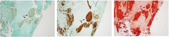
Chondrocytes and meniscal cells were isolated from articular cartilage and menisci of OA patients undergoing total knee arthroplasty. Chondrocyte- and meniscal cell-mediated calcification was examined using both monolayer and micromass culture-based assays. Crustal types were examined with histological staining. Levels of Type X Collagen, MMP-13, and ANKH in OA menisci were examined using immunohistochemistry.
Primary human OA meniscal cells produced calcified deposits at a similar rate compared to OA chondrocytes in-vitro. Histological examinations indicate that both BCP crystals and CPPD crystals are present in the meniscal tissue. Type X collagen, MMP-13, and ANKH were found in human OA menisci and their levels increased with OA severity. In addition, type X collagen was co-localized with calcium crystals.
These findings suggest that OA meniscal cells have a similar calcifying potential as OA chondrocytes, supporting a pathogenic role of OA menisci in OA.
传统上认为软骨细胞是骨关节炎膝关节内钙晶体沉积的原因。最近越来越多的实验证据表明半月板可能也起作用。然而,来自同一骨关节炎患者的软骨细胞和半月板细胞的钙化潜力以及与半月板钙化相关的基因从未得到充分研究。
研究并比较来自同一骨关节炎患者的关节软骨细胞和半月板细胞的钙化潜力,并确定骨关节炎半月板中的钙晶体类型和选定的基因表达。
从接受全膝关节置换术的骨关节炎患者的关节软骨和半月板中分离软骨细胞和半月板细胞。使用基于单层和微团培养的试验检测软骨细胞和半月板细胞介导的钙化。用组织学染色检查晶体类型。使用免疫组织化学检测骨关节炎半月板中X型胶原蛋白、基质金属蛋白酶-13(MMP-13)和ANKH的水平。
原代人骨关节炎半月板细胞在体外产生钙化沉积物的速率与骨关节炎软骨细胞相似。组织学检查表明半月板组织中同时存在羟基磷灰石晶体(BCP晶体)和焦磷酸钙晶体(CPPD晶体)。在人骨关节炎半月板中发现了X型胶原蛋白、MMP-13和ANKH,并且它们的水平随着骨关节炎严重程度的增加而升高。此外,X型胶原蛋白与钙晶体共定位。
这些发现表明骨关节炎半月板细胞具有与骨关节炎软骨细胞相似的钙化潜力,支持骨关节炎半月板在骨关节炎中的致病作用。