Meyer Franziska, Dittmann Annalena, Kornak Uwe, Herbster Maria, Pap Thomas, Lohmann Christoph H, Bertrand Jessica
Department of Orthopaedic Surgery, Otto-von-Guericke University Magdeburg, Magdeburg, Germany.
Institut für Humangenetik, Universitätsmedizin Göttingen, Göttingen, Germany.
Front Cell Dev Biol. 2021 Apr 26;9:622287. doi: 10.3389/fcell.2021.622287. eCollection 2021.
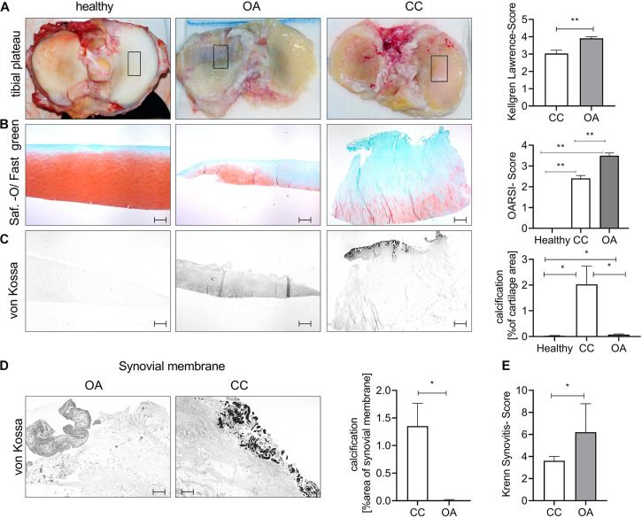
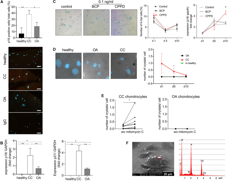
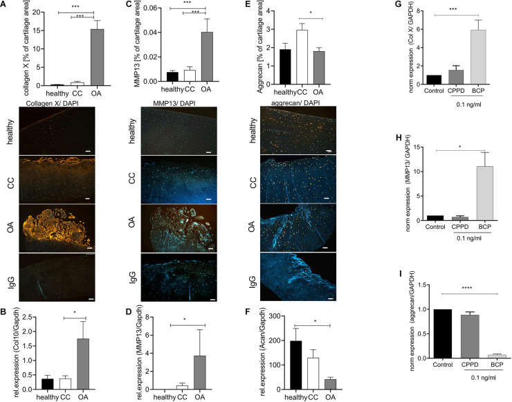
Basic calcium phosphate (BCP)-based calcification of cartilage is a common finding during osteoarthritis (OA) and is directly linked to the severity of the disease and hypertrophic differentiation of chondrocytes. Chondrocalcinosis (CC) is associated with calcium pyrophosphate dihydrate (CPPD) deposition disease in the joint inducing OA-like symptoms. There is only little knowledge about the effect of CPPD crystals on chondrocytes and the signaling pathways involved in their generation. The aim of this study was to investigate the chondrocyte phenotype in CC cartilage and the effect of CPPD crystals on chondrocytes. Cartilage samples of patients with CC, patients with severe OA, and healthy donors were included in this study. The presence of CC was evaluated using standard X-ray pictures, as well as von Kossa staining of cartilage sections. OA severity was evaluated using the Chambers Score on cartilage sections, as well as the radiological Kellgren-Lawrence Score. Patients with radiologically detectable CC presented calcification mainly on the cartilage surface, whereas OA patients showed calcification mainly in the pericellular matrix of hypertrophic chondrocytes. OA cartilage exhibited increased levels of collagen X and matrix metalloproteinase 13 (MMP13) compared with CC and healthy cartilage. This observation was confirmed by qRT-PCR using cartilage samples. No relevant influence of CPPD crystals on hypertrophic marker genes was observed , whereas BCP crystals significantly induced hypertrophic differentiation of chondrocytes. Interestingly, we observed an increased expression of p16 and p21 in cartilage samples of CC patients compared with OA patients and healthy controls, indicating cellular senescence. To investigate whether CPPD crystals were sufficient to induce senescence, we incubated chondrocytes with BCP and CPPD crystals and quantified senescence using β-gal staining. No significant difference was observed for the staining, but an increase of p16 expression was observed after 10 days of culture. Primary chondrocytes from CC patients produced CPPD crystals in culture. This phenotype was stabilized by mitomycin C-induced senescence. Healthy and OA chondrocytes did not exhibit this phenotype. BCP and CPPD crystals seem to be associated with two different chondrocyte phenotypes. Whereas BCP deposition is associated with chondrocyte hypertrophy, CPPD deposition is associated with cellular senescence.
基于碱性磷酸钙(BCP)的软骨钙化是骨关节炎(OA)期间的常见现象,并且与疾病的严重程度和软骨细胞的肥大分化直接相关。软骨钙质沉着症(CC)与关节中的焦磷酸钙二水合物(CPPD)沉积病相关,可诱发类似OA的症状。关于CPPD晶体对软骨细胞的影响及其生成所涉及的信号通路,人们了解甚少。本研究的目的是研究CC软骨中的软骨细胞表型以及CPPD晶体对软骨细胞的影响。本研究纳入了CC患者、重度OA患者和健康供体的软骨样本。使用标准X线片以及软骨切片的冯·科萨染色评估CC的存在情况。使用软骨切片上的钱伯斯评分以及放射学凯尔格伦-劳伦斯评分评估OA的严重程度。放射学上可检测到CC的患者钙化主要出现在软骨表面,而OA患者的钙化主要出现在肥大软骨细胞的细胞周围基质中。与CC和健康软骨相比,OA软骨中X型胶原蛋白和基质金属蛋白酶13(MMP13)的水平升高。使用软骨样本进行的qRT-PCR证实了这一观察结果。未观察到CPPD晶体对肥大标记基因有相关影响,而BCP晶体显著诱导软骨细胞肥大分化。有趣的是,我们观察到与OA患者和健康对照相比,CC患者软骨样本中p16和p21的表达增加,表明细胞衰老。为了研究CPPD晶体是否足以诱导衰老,我们用BCP和CPPD晶体培养软骨细胞,并使用β-半乳糖苷酶染色对衰老进行定量。染色未观察到显著差异,但培养10天后观察到p16表达增加。CC患者的原代软骨细胞在培养中产生CPPD晶体。丝裂霉素C诱导的衰老使这种表型稳定。健康和OA软骨细胞未表现出这种表型。BCP和CPPD晶体似乎与两种不同的软骨细胞表型相关。BCP沉积与软骨细胞肥大相关,而CPPD沉积与细胞衰老相关。