Haghiralsadat Fateme, Amoabediny Ghasem, Sheikhha Mohammad Hasan, Forouzanfar Tymour, Helder Marco N, Zandieh-Doulabi Behrouz
Department of Life Science Engineering, Faculty of New Sciences and Technologies, University of Tehran, Tehran, Iran.
Department of Nano Biotechnology, Research Center for New Technologies in Life Science Engineering, University of Tehran, Tehran, Iran.
Cell J. 2017 Spring;19(Suppl 1):55-65. doi: 10.22074/cellj.2017.4502. Epub 2017 May 17.
In this study we prepared a novel formulation of liposomal doxorubicin (L- DOX). The drug dose was optimized by analyses of cellular uptake and cell viability of osteosarcoma (OS) cell lines upon exposure to nanoliposomes that contained varying DOX concentrations. We intended to reduce the cytotoxicity of DOX and improve characteristics of the nanosystems.
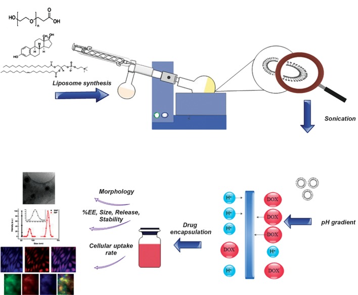
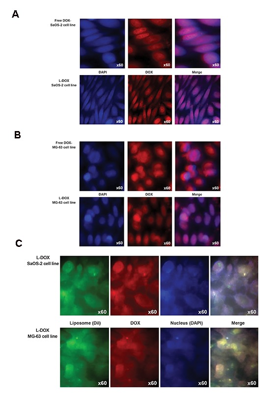
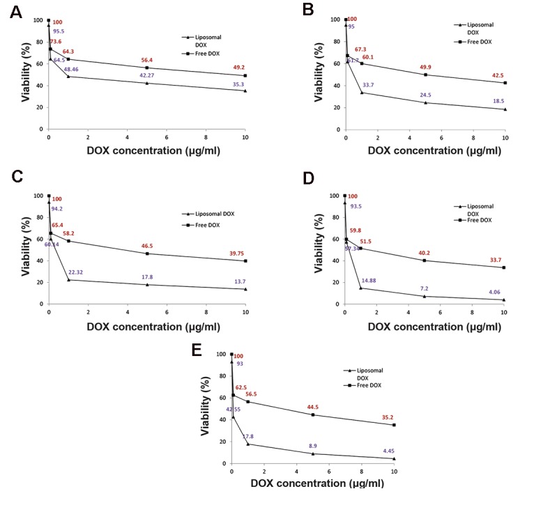
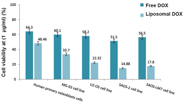
In this experimental study, we prepared liposomes by the pH gradient hydration method. Various characterization tests that included dynamic light scattering (DLS), cryogenic transmission electron microscopy (Cryo-TEM) imaging, and UV- Vis spectrophotometry were employed to evaluate the quality of the nanocarriers. In addition, the CyQUANT® assay and fluorescence microscope imaging were used on various OS cell lines (MG-63, U2-OS, SaOS-2, SaOS-LM7) and Human primary osteoblasts cells, as novel methods to determine cell viability and transfection efficacy.
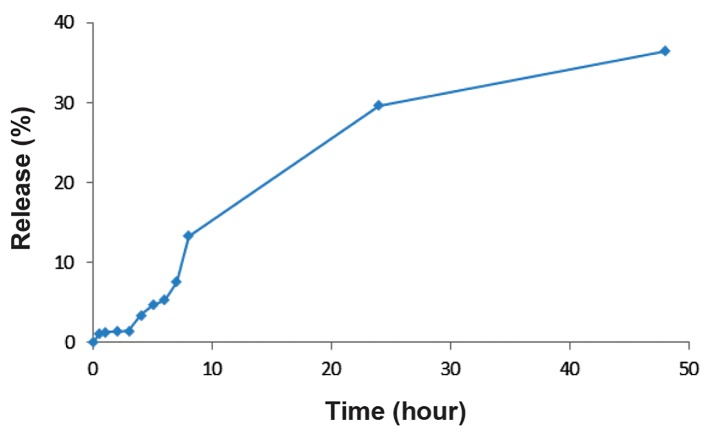
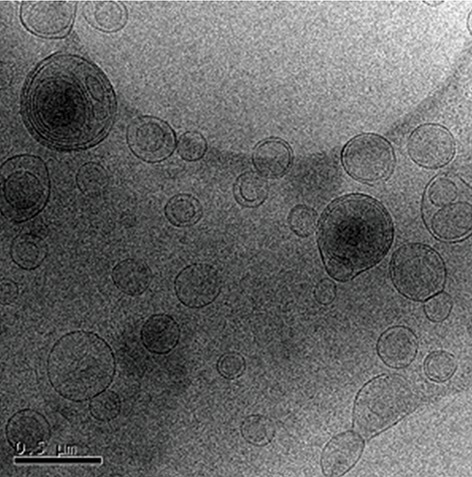
We observed an entrapment efficiency of 84% for DOX within the optimized liposomal formulation (L-DOX) that had a liposomal diameter of 96 nm. Less than 37% of DOX released after 48 hours and L-DOX could be stored stably for 14 days. L-DOX increased DOX toxicity by 1.8-4.6 times for the OS cell lines and only 1.3 times for Human primary osteoblasts cells compared to free DOX, which confirmed a higher sensitivity of the OS cell lines versus Human primary osteoblasts cells for L-DOX. We deduced that L- DOX passed more freely through the cell membrane compared to free DOX.
We successfully synthesized a stealth L-DOX that contained natural phospholipid by the pH gradient method, which could encapsulate DOX with 84% efficiency. The resulting nanoparticles were round, with a suitable particle size, and stable for 14 days. These nanoparticles allowed for adequately controlled DOX release, increased cell permeability compared to free DOX, and increased tumor cell death. L-DOX provided a novel, more effective therapy for OS treatment.
在本研究中,我们制备了一种新型的脂质体阿霉素(L-DOX)制剂。通过分析骨肉瘤(OS)细胞系在暴露于含有不同阿霉素浓度的纳米脂质体后的细胞摄取和细胞活力,对药物剂量进行了优化。我们旨在降低阿霉素的细胞毒性并改善纳米系统的特性。
在本实验研究中,我们采用pH梯度水化法制备脂质体。采用包括动态光散射(DLS)、低温透射电子显微镜(Cryo-TEM)成像和紫外可见分光光度法在内的各种表征测试来评估纳米载体的质量。此外,CyQUANT® 测定法和荧光显微镜成像被用于各种OS细胞系(MG-63、U2-OS、SaOS-2、SaOS-LM7)和人原代成骨细胞,作为确定细胞活力和转染效率的新方法。
我们观察到在优化的脂质体制剂(L-DOX)中阿霉素的包封率为84%,其脂质体直径为96 nm。48小时后释放的阿霉素不到37%,且L-DOX可稳定储存14天。与游离阿霉素相比,L-DOX使OS细胞系的阿霉素毒性增加了1.8至4.6倍,而对人原代成骨细胞仅增加了1.3倍,这证实了OS细胞系对L-DOX的敏感性高于人原代成骨细胞。我们推断,与游离阿霉素相比,L-DOX能更自由地穿过细胞膜。
我们通过pH梯度法成功合成了一种含有天然磷脂的隐形L-DOX,其能以84%的效率包封阿霉素。所得纳米颗粒呈圆形,粒径合适,且14天内稳定。这些纳米颗粒可实现对阿霉素释放的充分控制,与游离阿霉素相比增加了细胞通透性,并增加了肿瘤细胞死亡。L-DOX为OS治疗提供了一种新的、更有效的疗法。