• 文献检索
  • 文档翻译
  • 深度研究
  • 学术资讯
  • Suppr Zotero 插件Zotero 插件
  • 邀请有礼
  • 套餐&价格
  • 历史记录
应用&插件
Suppr Zotero 插件Zotero 插件浏览器插件Mac 客户端Windows 客户端微信小程序
定价
高级版会员购买积分包购买API积分包
服务
文献检索文档翻译深度研究API 文档MCP 服务
关于我们
关于 Suppr公司介绍联系我们用户协议隐私条款
关注我们

Suppr 超能文献

核心技术专利:CN118964589B侵权必究
粤ICP备2023148730 号-1Suppr @ 2026

文献检索

告别复杂PubMed语法,用中文像聊天一样搜索,搜遍4000万医学文献。AI智能推荐,让科研检索更轻松。

立即免费搜索

文件翻译

保留排版,准确专业,支持PDF/Word/PPT等文件格式,支持 12+语言互译。

免费翻译文档

深度研究

AI帮你快速写综述,25分钟生成高质量综述,智能提取关键信息,辅助科研写作。

立即免费体验

乳腺体积密度对筛查发现的乳腺癌和间期乳腺癌风险的影响:一项队列研究。

The effect of volumetric breast density on the risk of screen-detected and interval breast cancers: a cohort study.

作者信息

Wanders Johanna O P, Holland Katharina, Karssemeijer Nico, Peeters Petra H M, Veldhuis Wouter B, Mann Ritse M, van Gils Carla H

机构信息

Julius Center for Health Sciences and Primary Care, University Medical Center Utrecht, P.O. Box 85500, 3508 GA, Utrecht, The Netherlands.

Department of Radiology and Nuclear Medicine, Radboud University Medical Center, Geert Grooteplein 10, 6525 GA, Nijmegen, The Netherlands.

出版信息

Breast Cancer Res. 2017 Jun 5;19(1):67. doi: 10.1186/s13058-017-0859-9.

DOI:10.1186/s13058-017-0859-9
PMID:28583146
原文链接:https://pmc.ncbi.nlm.nih.gov/articles/PMC5460501/
Abstract

BACKGROUND

In the light of the breast density legislation in the USA, it is important to know a woman's breast cancer risk, but particularly her risk of a tumor that is not detected through mammographic screening (interval cancer). Therefore, we examined the associations of automatically measured volumetric breast density with screen-detected and interval cancer risk, separately.

METHODS

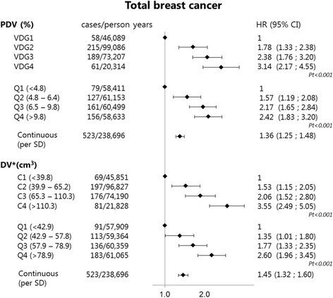
Volumetric breast measures were assessed automatically using Volpara version 1.5.0 (Matakina, New Zealand) for the first available digital mammography (DM) examination of 52,814 women (age 50 - 75 years) participating in the Dutch biennial breast cancer screening program between 2003 and 2011. Breast cancer information was obtained from the screening registration system and through linkage with the Netherlands Cancer Registry. We excluded all screen-detected breast cancers diagnosed as a result of the first digital screening examination. During a median follow-up period of 4.2 (IQR 2.0-6.2) years, 523 women were diagnosed with breast cancer of which 299 were screen-detected and 224 were interval breast cancers. The associations between volumetric breast measures and breast cancer risk were determined using Cox proportional hazards analyses.

RESULTS

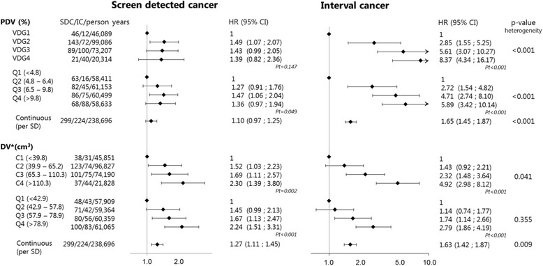
Percentage dense volume was found to be positively associated with both interval and screen-detected breast cancers (hazard ratio (HR) 8.37 (95% CI 4.34-16.17) and HR 1.39 (95% CI 0.82-2.36), respectively, for Volpara density grade category (VDG) 4 compared to VDG1 (p for heterogeneity < 0.001)). Dense volume (DV) was also found to be positively associated with both interval and screen-detected breast cancers (HR 4.92 (95% CI 2.98-8.12) and HR 2.30 (95% CI 1.39-3.80), respectively, for VDG-like category (C)4 compared to C1 (p for heterogeneity = 0.041)). The association between percentage dense volume categories and interval breast cancer risk (HR 8.37) was not significantly stronger than the association between absolute dense volume categories and interval breast cancer risk (HR 4.92).

CONCLUSIONS

Our results suggest that both absolute dense volume and percentage dense volume are strong markers of breast cancer risk, but that they are even stronger markers for predicting the occurrence of tumors that are not detected during mammography breast cancer screening.

摘要

背景

鉴于美国的乳房密度立法,了解女性患乳腺癌的风险很重要,尤其是她患通过乳房X线筛查未被发现的肿瘤(间期癌)的风险。因此,我们分别研究了自动测量的乳房体积密度与筛查发现的癌症和间期癌风险之间的关联。

方法

使用Volpara 1.5.0版本(新西兰Matakina公司)对参与2003年至2011年荷兰两年一次乳腺癌筛查项目的52814名女性(年龄50 - 75岁)的首次可用数字化乳腺摄影(DM)检查进行乳房体积测量的自动评估。乳腺癌信息从筛查登记系统获取,并通过与荷兰癌症登记处的链接获得。我们排除了因首次数字化筛查检查而诊断出的所有筛查发现的乳腺癌。在中位随访期4.2(四分位间距2.0 - 6.2)年期间,523名女性被诊断患有乳腺癌,其中299例为筛查发现,224例为间期乳腺癌。使用Cox比例风险分析确定乳房体积测量与乳腺癌风险之间的关联。

结果

发现致密体积百分比与间期癌和筛查发现的乳腺癌均呈正相关(与VDG1相比,Volpara密度等级类别(VDG)4的风险比(HR)分别为8.37(95%置信区间4.34 - 16.17)和HR 1.39(95%置信区间0.82 - 2.36),异质性p < 0.001)。致密体积(DV)也被发现与间期癌和筛查发现的乳腺癌均呈正相关(与C1相比,VDG样类别(C)4的HR分别为4.92(95%置信区间2.98 - 8.12)和HR 2.30(95%置信区间1.39 - 3.80),异质性p = 0.041)。致密体积百分比类别与间期乳腺癌风险之间的关联(HR 8.37)并不比绝对致密体积类别与间期乳腺癌风险之间的关联(HR 4.9)显著更强。

结论

我们的结果表明,绝对致密体积和致密体积百分比都是乳腺癌风险的有力标志物,但它们更是预测在乳房X线乳腺癌筛查期间未被发现的肿瘤发生的更强标志物。

https://cdn.ncbi.nlm.nih.gov/pmc/blobs/4187/5460501/b7b3732992e3/13058_2017_859_Fig2_HTML.jpg
https://cdn.ncbi.nlm.nih.gov/pmc/blobs/4187/5460501/dc560dec6af9/13058_2017_859_Fig1_HTML.jpg
https://cdn.ncbi.nlm.nih.gov/pmc/blobs/4187/5460501/b7b3732992e3/13058_2017_859_Fig2_HTML.jpg
https://cdn.ncbi.nlm.nih.gov/pmc/blobs/4187/5460501/dc560dec6af9/13058_2017_859_Fig1_HTML.jpg
https://cdn.ncbi.nlm.nih.gov/pmc/blobs/4187/5460501/b7b3732992e3/13058_2017_859_Fig2_HTML.jpg

相似文献

1
The effect of volumetric breast density on the risk of screen-detected and interval breast cancers: a cohort study.乳腺体积密度对筛查发现的乳腺癌和间期乳腺癌风险的影响:一项队列研究。
Breast Cancer Res. 2017 Jun 5;19(1):67. doi: 10.1186/s13058-017-0859-9.
2
The combined effect of mammographic texture and density on breast cancer risk: a cohort study.乳腺影像纹理与密度联合对乳腺癌风险的影响:一项队列研究。
Breast Cancer Res. 2018 May 2;20(1):36. doi: 10.1186/s13058-018-0961-7.
3
Volumetric breast density affects performance of digital screening mammography.乳腺体积密度会影响数字化乳腺筛查钼靶摄影的效果。
Breast Cancer Res Treat. 2017 Feb;162(1):95-103. doi: 10.1007/s10549-016-4090-7. Epub 2016 Dec 23.
4
Automated measurement of volumetric mammographic density: a tool for widespread breast cancer risk assessment.乳腺钼靶体积密度的自动化测量:一种广泛用于乳腺癌风险评估的工具。
Cancer Epidemiol Biomarkers Prev. 2014 Sep;23(9):1764-72. doi: 10.1158/1055-9965.EPI-13-1219. Epub 2014 Jul 10.
5
AI performance by mammographic density in a retrospective cohort study of 99,489 participants in BreastScreen Norway.挪威乳腺筛查项目中 99489 名参与者的回顾性队列研究中乳腺密度对 AI 性能的影响。
Eur Radiol. 2024 Oct;34(10):6298-6308. doi: 10.1007/s00330-024-10681-z. Epub 2024 Mar 25.
6
Breast Cancer Risk and Mammographic Density Assessed with Semiautomated and Fully Automated Methods and BI-RADS.采用半自动和全自动方法及乳腺影像报告和数据系统(BI-RADS)评估乳腺癌风险和乳腺钼靶密度
Radiology. 2017 Feb;282(2):348-355. doi: 10.1148/radiol.2016152062. Epub 2016 Sep 5.
7
Breast density and breast cancer-specific survival by detection mode.按检测方式划分的乳腺密度与乳腺癌特异性生存
BMC Cancer. 2018 Apr 5;18(1):386. doi: 10.1186/s12885-018-4316-7.
8
Novel mammographic image features differentiate between interval and screen-detected breast cancer: a case-case study.新型乳腺钼靶图像特征可区分间期癌和筛查发现的乳腺癌:一项病例对照研究。
Breast Cancer Res. 2016 Oct 5;18(1):100. doi: 10.1186/s13058-016-0761-x.
9
Digital versus screen-film mammography: impact of mammographic density and hormone therapy on breast cancer detection.数字化乳腺钼靶摄影与屏-片乳腺钼靶摄影:乳腺密度和激素治疗对乳腺癌检测的影响
Breast Cancer Res Treat. 2015 Nov;154(2):377-87. doi: 10.1007/s10549-015-3622-x. Epub 2015 Oct 30.
10
Digital volumetric measurement of mammographic density and the risk of overlooking cancer in Japanese women.日本女性乳腺钼靶密度的数字容积测量及癌症漏诊风险
Breast Cancer. 2017 Sep;24(5):708-713. doi: 10.1007/s12282-017-0763-2. Epub 2017 Feb 25.

引用本文的文献

1
Machine learning-based classification model to differentiate subtypes of invasive breast cancer using MRI.基于机器学习的分类模型,用于利用磁共振成像鉴别浸润性乳腺癌的亚型
Front Oncol. 2025 Jun 3;15:1588787. doi: 10.3389/fonc.2025.1588787. eCollection 2025.
2
Mammographic density by time and breast: a retrospective cohort study from BreastScreen Norway.不同时间及不同乳房的乳腺钼靶密度:来自挪威乳腺筛查项目的一项回顾性队列研究
Breast Cancer Res. 2025 May 16;27(1):83. doi: 10.1186/s13058-025-02037-2.
3
A practical work around for breast density distribution discrepancies between mammographic images from different vendors.

本文引用的文献

1
Volumetric breast density affects performance of digital screening mammography.乳腺体积密度会影响数字化乳腺筛查钼靶摄影的效果。
Breast Cancer Res Treat. 2017 Feb;162(1):95-103. doi: 10.1007/s10549-016-4090-7. Epub 2016 Dec 23.
2
Breast Cancer Risk and Mammographic Density Assessed with Semiautomated and Fully Automated Methods and BI-RADS.采用半自动和全自动方法及乳腺影像报告和数据系统(BI-RADS)评估乳腺癌风险和乳腺钼靶密度
Radiology. 2017 Feb;282(2):348-355. doi: 10.1148/radiol.2016152062. Epub 2016 Sep 5.
3
Variation in Mammographic Breast Density Assessments Among Radiologists in Clinical Practice: A Multicenter Observational Study.
一种解决不同供应商乳腺钼靶图像之间乳腺密度分布差异的实用方法。
Eur Radiol. 2025 Jan 31. doi: 10.1007/s00330-025-11383-w.
4
Diagnostic Accuracy of Screening Contrast-enhanced Mammography for Women with Extremely Dense Breasts at Increased Risk of Breast Cancer.筛查对比增强乳腺摄影对乳腺癌高危致密乳腺女性的诊断准确性。
Radiology. 2024 Oct;313(1):e232580. doi: 10.1148/radiol.232580.
5
Awareness of Breast Cancer Risk Factors in Women with vs. Without High Breast Density.有高乳腺密度与无高乳腺密度女性对乳腺癌风险因素的认知情况。
Patient Prefer Adherence. 2024 Jul 31;18:1577-1588. doi: 10.2147/PPA.S466992. eCollection 2024.
6
[Results of Lung Cancer Screening with Low-dose Computed Tomography 
and Exploration of Risk Factors in Guangzhou].[广州地区低剂量计算机断层扫描肺癌筛查结果及危险因素探索]
Zhongguo Fei Ai Za Zhi. 2024 May 20;27(5):345-358. doi: 10.3779/j.issn.1009-3419.2024.101.14.
7
Influence of mammographic density and compressed breast thickness on true mammographic sensitivity: a cohort study.乳腺密度和压缩乳房厚度对真实乳腺钼靶灵敏度的影响:一项队列研究。
Sci Rep. 2023 Aug 30;13(1):14194. doi: 10.1038/s41598-023-41356-2.
8
Clinical and imaging modality factors impacting radiological interpretation of breast screening in young women with neurofibromatosis type 1.影响 1 型神经纤维瘤病年轻女性乳腺筛查影像学解读的临床和影像学因素。
Fam Cancer. 2023 Oct;22(4):499-511. doi: 10.1007/s10689-023-00340-5. Epub 2023 Jun 19.
9
Impact of BMI on Prevalence of Dense Breasts by Race and Ethnicity.体质量指数(BMI)对不同种族和民族致密型乳腺患病率的影响。
Cancer Epidemiol Biomarkers Prev. 2023 Nov 1;32(11):1524-1530. doi: 10.1158/1055-9965.EPI-23-0049.
10
Multi-vendor robustness analysis of a commercial artificial intelligence system for breast cancer detection.用于乳腺癌检测的商用人工智能系统的多供应商稳健性分析。
J Med Imaging (Bellingham). 2023 Sep;10(5):051807. doi: 10.1117/1.JMI.10.5.051807. Epub 2023 Apr 18.
临床实践中放射科医生对乳腺钼靶密度评估的差异:一项多中心观察性研究。
Ann Intern Med. 2016 Oct 4;165(7):457-464. doi: 10.7326/M15-2934. Epub 2016 Jul 19.
4
Mammographic density and risk of breast cancer by mode of detection and tumor size: a case-control study.根据检测方式和肿瘤大小分析乳腺钼靶密度与乳腺癌风险:一项病例对照研究。
Breast Cancer Res. 2016 Jun 18;18(1):63. doi: 10.1186/s13058-016-0722-4.
5
Comparison of Clinical and Automated Breast Density Measurements: Implications for Risk Prediction and Supplemental Screening.临床与自动乳腺密度测量的比较:对风险预测和补充筛查的意义。
Radiology. 2016 Jun;279(3):710-9. doi: 10.1148/radiol.2015151261. Epub 2015 Dec 22.
6
Postmenopausal mammographic breast density and subsequent breast cancer risk according to selected tissue markers.根据选定的组织标志物评估绝经后乳腺钼靶密度与后续乳腺癌风险
Br J Cancer. 2015 Sep 29;113(7):1104-13. doi: 10.1038/bjc.2015.315. Epub 2015 Sep 3.
7
Misclassification of Breast Imaging Reporting and Data System (BI-RADS) Mammographic Density and Implications for Breast Density Reporting Legislation.乳腺影像报告和数据系统(BI-RADS)中乳腺钼靶密度的错误分类及其对乳腺密度报告立法的影响
Breast J. 2015 Sep-Oct;21(5):481-9. doi: 10.1111/tbj.12443. Epub 2015 Jul 1.
8
Identifying women with dense breasts at high risk for interval cancer: a cohort study.识别患间期癌风险高的致密乳腺女性:一项队列研究。
Ann Intern Med. 2015 May 19;162(10):673-81. doi: 10.7326/M14-1465.
9
Risk factors and tumor characteristics of interval cancers by mammographic density.基于乳腺密度的间隔期癌症的风险因素和肿瘤特征。
J Clin Oncol. 2015 Mar 20;33(9):1030-7. doi: 10.1200/JCO.2014.58.9986. Epub 2015 Feb 2.
10
Mammographic features associated with interval breast cancers in screening programs.筛查项目中与间期乳腺癌相关的乳腺钼靶特征。
Breast Cancer Res. 2014 Aug 26;16(4):417. doi: 10.1186/s13058-014-0417-7.