Peca Stefano, Brown Derek Wilson, Smith Wendy Lani
Department of Physics and Astronomy, University of Calgary, Calgary, AB, Canada.
Department of Medical Physics, Tom Baker Cancer Centre, Calgary, AB, Canada.
Technol Cancer Res Treat. 2017 Dec;16(6):944-955. doi: 10.1177/1533034617711354. Epub 2017 Jun 6.
To improve patient safety and treatment quality, verification of dose delivery in radiotherapy is desirable. We present a simple, easy-to-implement, open-source method for planar dosimetry of conformal radiotherapy by electronic portal imaging device (EPID).
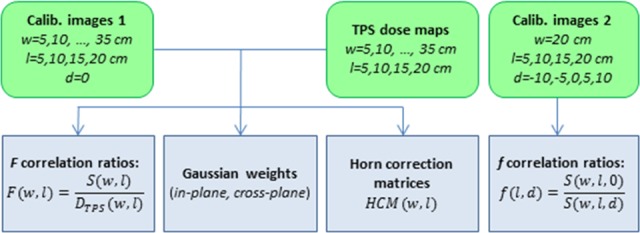
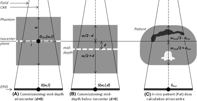
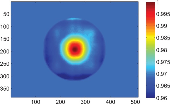
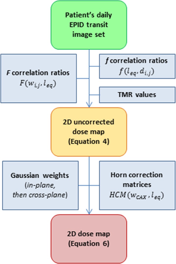
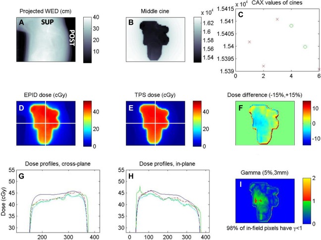
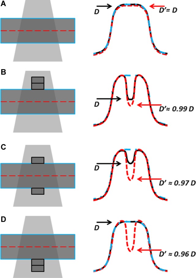
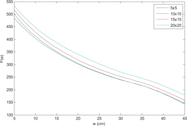
Correlation ratios, which relate dose in the mid-depth of slab phantoms to transit EPID signal, were determined for multiple phantom thicknesses and field sizes. Off-axis dose is corrected for by means of model-based convolution. We tested efficacy of dose reconstruction through measurements with off-reference values of attenuator thickness, field size, and monitor units. We quantified the dose calculation error in the presence of thickness changes to simulate anatomical or setup variations. An example of dose calculation on patient data is provided.
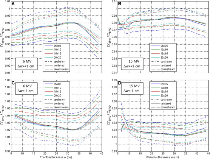
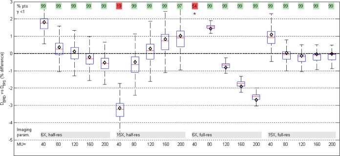
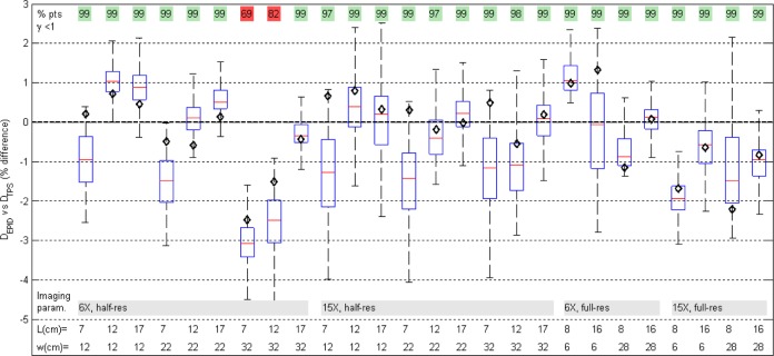
With varying phantom thickness, field size, and monitor units, dose reconstruction was almost always within 3% of planned dose. In the presence of thickness changes from planning CT, the dose discrepancy is exaggerated by up to approximately 1.5% for 1 cm changes upstream of the isocenter plane and 4% for 1 cm changes downstream.
Our novel electronic portal imaging device dosimetry allows clinically accurate 2-dimensional reconstruction of dose inside a phantom/patient at isocenter depth. Due to its simplicity, commissioning can be performed in a few hours per energy and may be modified to the user's needs. It may provide useful dose delivery information to detect harmful errors, guide adaptive radiotherapy, and assure quality of treatment.
为提高患者安全和治疗质量,放疗中剂量输送的验证是很有必要的。我们提出了一种简单、易于实施的开源方法,用于通过电子射野影像装置(EPID)对适形放疗进行平面剂量测定。
针对多种体模厚度和射野尺寸,确定了平板体模中深度处的剂量与传输的EPID信号之间的相关比。通过基于模型的卷积对离轴剂量进行校正。我们通过使用衰减器厚度、射野尺寸和监测单位的非参考值进行测量,测试了剂量重建的有效性。我们对存在厚度变化时的剂量计算误差进行了量化,以模拟解剖结构或摆位的变化。提供了一个患者数据剂量计算的示例。
随着体模厚度、射野尺寸和监测单位的变化,剂量重建几乎总是在计划剂量的3%以内。在存在与计划CT相比厚度变化的情况下,对于等中心平面上游1 cm的变化,剂量差异会被夸大至约1.5%,对于下游1 cm的变化,剂量差异会被夸大至4%。
我们新颖的电子射野影像装置剂量测定法能够在等中心深度对体模/患者体内剂量进行临床准确的二维重建。由于其简单性,每个能量的调试可在几小时内完成,并且可以根据用户需求进行修改。它可以提供有用的剂量输送信息,以检测有害误差、指导自适应放疗并确保治疗质量。