Zhang Peng, Zhi Yunlong, Fang Hongwei, Wu Ziying, Chen Tianwu, Jiang Jia, Chen Shiyi
Department of Sports Medicine, Huashan Hospital, Fudan University, Shanghai 200040, P.R. China.
Department of Anesthesiology, Bengbu Medical College, Bengbu, Anhui 233004, P.R. China.
Exp Ther Med. 2017 Jun;13(6):2751-2756. doi: 10.3892/etm.2017.4359. Epub 2017 Apr 19.
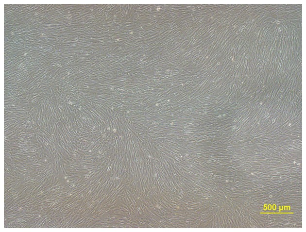
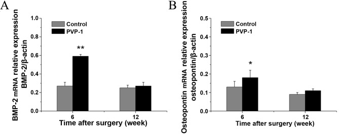
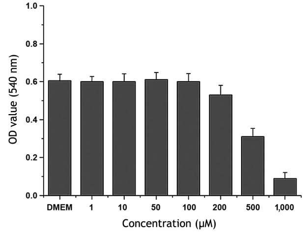
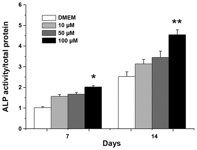
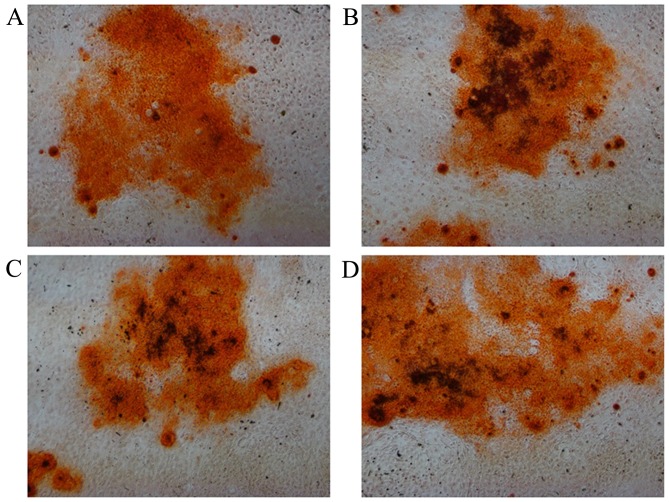
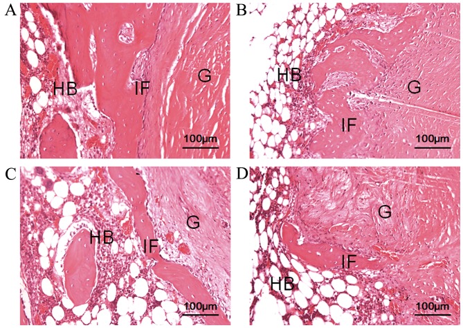
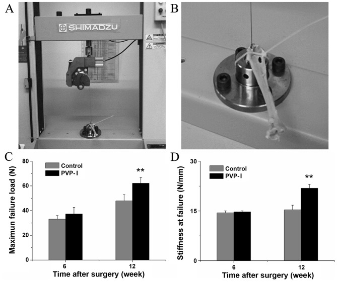
Polyvinylpyrrolidone-iodine (PVP-I) is a broad-spectrum antimicrobial agent, but its effects on tendon-bone healing are unclear. The purpose of this study was to investigate the effects of PVP-I on bone marrow mesenchymal stem cells (BMSCs) and on tendon-bone healing . In this study, following investigation of the concentration-dependent effects of PVP-I on the viability and osteogenic differentiation of BMSCs, the appropriate concentration of PVP-I was selected for animal experiments. New Zealand white rabbits received autologous tendon transplantation with and without PVP-I treatment of the graft tendon. Subsequently, histological examination, biomechanical testing and reverse transcription-quantitative polymerase chain reaction (RT-qPCR) analyses were conducted. At 6 weeks post-surgery, connective tissue and osteogenesis was observed at the tendon-bone interface in the PVP-I group. At 12 weeks post-surgery, the interface width in the PVP-I group was much narrower compared with that of the control group. Furthermore, the biomechanical properties of the PVP-I group were significantly stronger than those in the control group (P<0.05). RT-qPCR examination revealed that the mRNA levels of bone morphogenetic protein-2 and osteopontin in the PVP-I group were higher than those in the control group at 6 weeks (P<0.05). In conclusion, these results indicated that PVP-I promoted tendon-bone healing via osteogenesis.
聚维酮碘(PVP-I)是一种广谱抗菌剂,但其对肌腱-骨愈合的影响尚不清楚。本研究的目的是探讨PVP-I对骨髓间充质干细胞(BMSCs)以及肌腱-骨愈合的影响。在本研究中,在研究PVP-I对BMSCs活力和成骨分化的浓度依赖性影响后,选择合适浓度的PVP-I进行动物实验。新西兰白兔接受自体肌腱移植,移植的肌腱分别进行PVP-I处理和不进行PVP-I处理。随后,进行组织学检查、生物力学测试和逆转录定量聚合酶链反应(RT-qPCR)分析。术后6周,PVP-I组的肌腱-骨界面处观察到结缔组织和成骨现象。术后12周,PVP-I组的界面宽度比对照组窄得多。此外,PVP-I组的生物力学性能明显强于对照组(P<0.05)。RT-qPCR检测显示,PVP-I组在术后6周时骨形态发生蛋白-2和骨桥蛋白的mRNA水平高于对照组(P<0.05)。总之,这些结果表明PVP-I通过成骨作用促进了肌腱-骨愈合。