Department of Orthopedic Surgery, Zhongnan Hospital of Wuhan University, Wuhan, 430071, China.
Department of Pharmacology, Basic Medical School of Wuhan University, Wuhan, 430071, China.
Stem Cell Res Ther. 2021 Nov 14;12(1):576. doi: 10.1186/s13287-021-02643-9.
Bone formation plays an important role in early tendon-bone healing after anterior cruciate ligament reconstruction (ACLR). Dedifferentiated osteogenic bone marrow mesenchymal stem cells (De-BMSCs) have enhanced osteogenic potential. This study aimed to investigate the effect of De-BMSCs transplantation on the promotion of bone formation at the tendon-bone interface after ACLR and to further explore the molecular mechanism of the enhanced osteogenic potential of De-BMSCs.
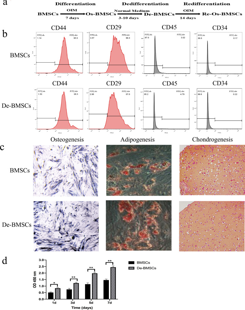
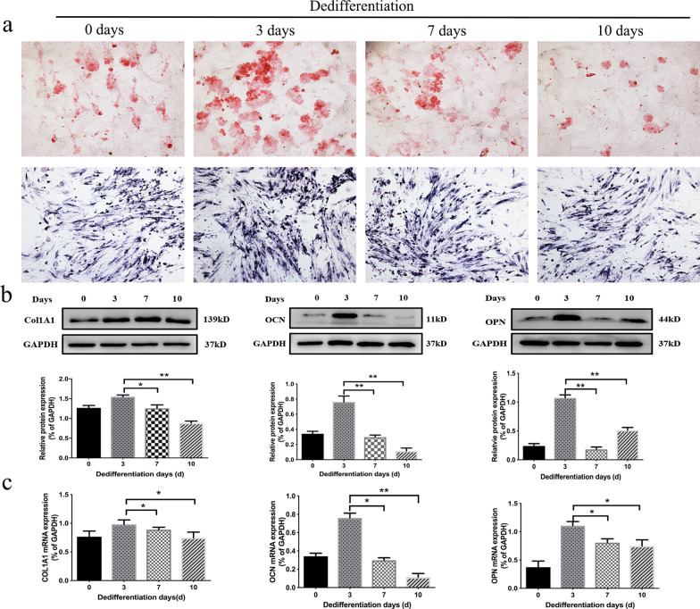
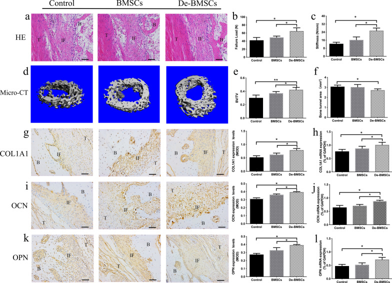
BMSCs from the femurs and tibias of New Zealand white rabbits were subjected to osteogenic induction and then cultured in medium without osteogenic factors; the obtained cell population was termed De-BMSCs. De-BMSCs were induced to undergo osteo-, chondro- and adipo-differentiation in vitro to examine the characteristics of primitive stem cells. An ACLR model with a semitendinosus tendon was established in rabbits, and the animals were divided into a control group, BMSCs group, and De-BMSCs group. At 12 weeks after surgery, the rabbits in each group were sacrificed to evaluate tendon-bone healing by histologic staining, micro-computed tomography (micro-CT) examination, and biomechanical testing. During osteogenic differentiation of De-BMSCs, an siRNA targeting nuclear factor of activated T-cells 1 (NFATc1) was used to verify the molecular mechanism of the enhanced osteogenic potential of De-BMSCs.
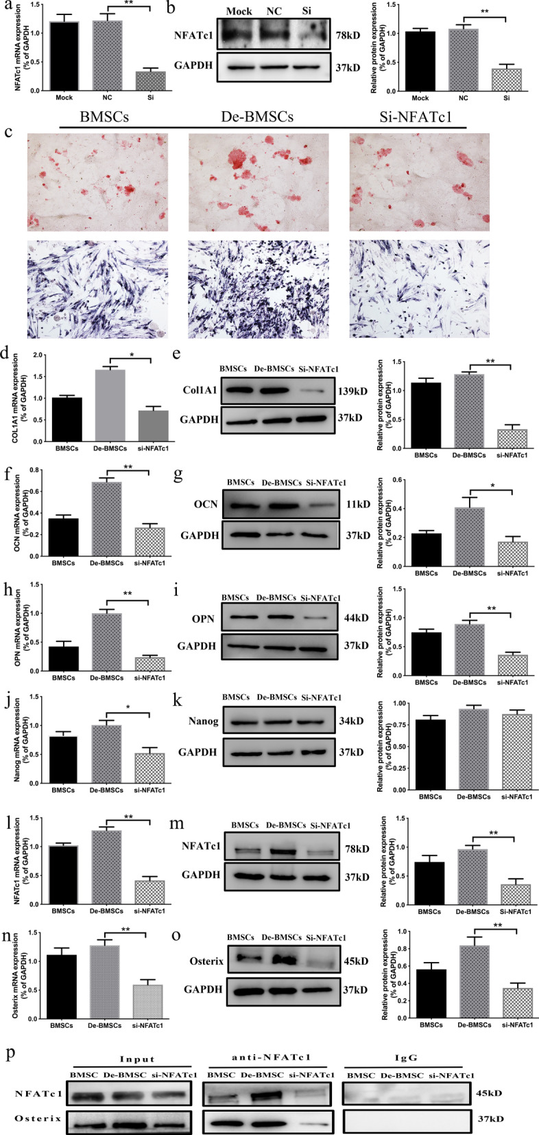
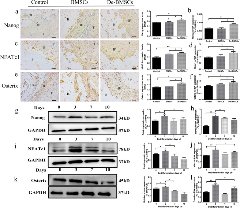
De-BMSCs exhibited some properties similar to BMSCs, including multiple differentiation potential and cell surface markers. Bone formation at the tendon-bone interface in the De-BMSCs group was significantly increased, and biomechanical strength was significantly improved. During the osteogenic differentiation of De-BMSCs, the expression of Nanog and NFATc1 was synergistically increased, which promoted the interaction of NFATc1 and Osterix, resulting in increased expression of osteoblast marker genes such as COL1A, OCN, and OPN.
De-BMSCs transplantation could promote bone formation at the tendon-bone interface after ACLR and improve the biomechanical strength of the reconstruction. The Nanog/NFATc1/Osterix signaling pathway mediated the enhanced osteogenic differentiation efficiency of De-BMSCs.
在前交叉韧带重建(ACLR)后,骨形成在早期腱骨愈合中起着重要作用。去分化成骨骨髓间充质干细胞(De-BMSCs)具有增强的成骨潜能。本研究旨在探讨 De-BMSCs 移植对 ACLR 后腱骨界面骨形成的促进作用,并进一步探讨 De-BMSCs 增强成骨潜能的分子机制。
从新西兰白兔的股骨和胫骨中提取 BMSCs,进行成骨诱导,然后在无成骨因子的培养基中培养;获得的细胞群体称为 De-BMSCs。体外诱导 De-BMSCs 向成骨、软骨和成脂分化,以鉴定原始干细胞的特征。在兔中建立 ACLR 模型(使用半腱肌腱),将动物分为对照组、BMSCs 组和 De-BMSCs 组。术后 12 周时,每组处死兔子,通过组织学染色、微计算机断层扫描(micro-CT)检查和生物力学测试评估腱骨愈合情况。在 De-BMSCs 的成骨分化过程中,使用针对活化 T 细胞核因子 1(NFATc1)的 siRNA 来验证 De-BMSCs 增强成骨潜能的分子机制。
De-BMSCs 表现出一些与 BMSCs 相似的特性,包括多向分化潜能和细胞表面标志物。De-BMSCs 组腱骨界面的骨形成明显增加,生物力学强度显著提高。在 De-BMSCs 的成骨分化过程中,Nanog 和 NFATc1 的表达协同增加,促进了 NFATc1 和 Osterix 的相互作用,导致成骨标记基因如 COL1A、OCN 和 OPN 的表达增加。
De-BMSCs 移植可促进 ACLR 后腱骨界面的骨形成,并提高重建的生物力学强度。Nanog/NFATc1/Osterix 信号通路介导了 De-BMSCs 增强的成骨分化效率。