• 文献检索
  • 文档翻译
  • 深度研究
  • 学术资讯
  • Suppr Zotero 插件Zotero 插件
  • 邀请有礼
  • 套餐&价格
  • 历史记录
应用&插件
Suppr Zotero 插件Zotero 插件浏览器插件Mac 客户端Windows 客户端微信小程序
定价
高级版会员购买积分包购买API积分包
服务
文献检索文档翻译深度研究API 文档MCP 服务
关于我们
关于 Suppr公司介绍联系我们用户协议隐私条款
关注我们

Suppr 超能文献

核心技术专利:CN118964589B侵权必究
粤ICP备2023148730 号-1Suppr @ 2026

文献检索

告别复杂PubMed语法,用中文像聊天一样搜索,搜遍4000万医学文献。AI智能推荐,让科研检索更轻松。

立即免费搜索

文件翻译

保留排版,准确专业,支持PDF/Word/PPT等文件格式,支持 12+语言互译。

免费翻译文档

深度研究

AI帮你快速写综述,25分钟生成高质量综述,智能提取关键信息,辅助科研写作。

立即免费体验

联动彩色成像提高结直肠息肉的可视性:一项视频研究。

Linked color imaging improves the visibility of colorectal polyps: a video study.

作者信息

Yoshida Naohisa, Naito Yuji, Murakami Takaaki, Hirose Ryohei, Ogiso Kiyoshi, Inada Yutaka, Dohi Osamu, Kamada Kazuhiro, Uchiyama Kazuhiko, Handa Osamu, Konishi Hideyuki, Siah Kewin Tien Ho, Yagi Nobuaki, Fujita Yasuko, Kishimoto Mitsuo, Yanagisawa Akio, Itoh Yoshito

机构信息

Department of Molecular Gastroenterology and Hepatology, Kyoto Prefectural University of Medicine, Graduate School of Medical Science, Kyoto, Japan.

Division of Gastroenterology & Hepatology, University Medicine Cluster, National University Hospital, Singapore.

出版信息

Endosc Int Open. 2017 Jun;5(6):E518-E525. doi: 10.1055/s-0043-105495. Epub 2017 Jun 7.

DOI:10.1055/s-0043-105495
PMID:28596985
原文链接:https://pmc.ncbi.nlm.nih.gov/articles/PMC5462607/
Abstract

BACKGROUND/STUDY AIM: Linked color imaging (LCI) by a laser endoscope (Fujifilm Co, Tokyo, Japan) is a novel narrow band light observation. In this study, we aimed to investigate whether LCI could improve the visibility of colorectal polyps using endoscopic videos.

PATIENTS AND METHODS

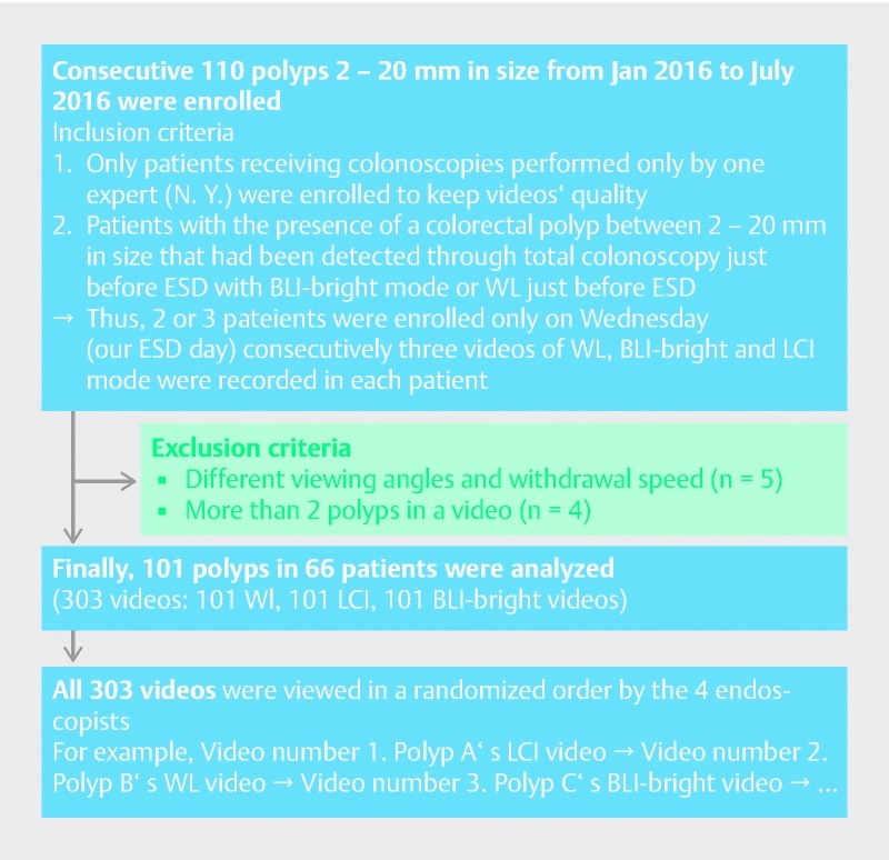
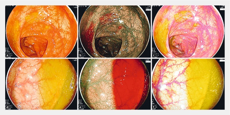
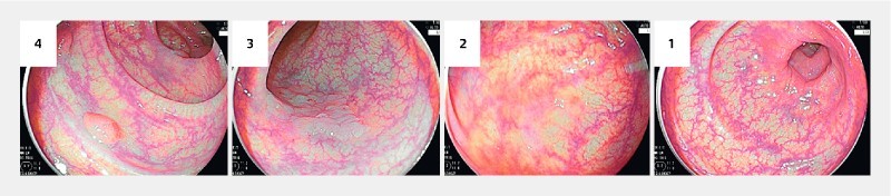
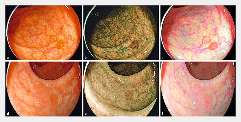
We prospectively recorded videos of consecutive polyps 2 - 20 mm in size diagnosed as neoplastic polyps. Three videos, white light (WL), blue laser imaging (BLI)-bright, and LCI, were recorded for each polyp by one expert. After excluding inappropriate videos, all videos were evaluated in random order by two experts and two non-experts according to a published polyp visibility score from four (excellent visibility) to one (poor visibility). Additionally, the relationship between polyp visibility scores in LCI and various clinical characteristics including location, size, histology, morphology, and preparation were analyzed compared to WL and BLI-bright.

RESULTS

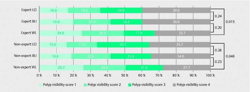
We analyzed 101 colorectal polyps (94 neoplastic) in 66 patients (303 videos). The mean polyp size was 9.0 ± 8.1 mm and 54 polyps were non-polypoid. The mean polyp visibility scores for LCI (2.86 ± 1.08) were significantly higher than for WL and BLI-bright (2.53 ± 1.15,  < 0.001; 2.73 ± 1.47,  < 0.041). The ratio of poor visibility (score 1 and 2) was significantly lower in LCI for experts and non-experts (35.6 %, 33.6 %) compared with WL (49.6 %,  = 0.015, 50.5 %,  = 0.046). The polyp visibility scores for LCI were significantly higher than those for WL for all of the factors. With respect to the comparison between BLI-bright and WL, the polyp visibility scores for BLI-bright were not higher than WL for right-sided location, < 10 mm size, sessile serrated adenoma and polyp histology, and poor preparation. For those characteristics, LCI improved the lesions with right-sided location, SSA/P histology, and poor preparation significantly better than BLI.

CONCLUSIONS

LCI improved polyp visibility compared to WL for both expert and non-expert endoscopists. It is useful for improving polyp visibility in any location, any size, any morphology, any histology, and any preparation level.

摘要

背景/研究目的:使用激光内镜(日本东京富士胶片公司)进行的联动成像(LCI)是一种新型窄带光观察技术。在本研究中,我们旨在通过内镜视频研究LCI是否能提高大肠息肉的可视性。

患者与方法

我们前瞻性地记录了连续诊断为肿瘤性息肉、大小在2 - 20毫米之间的息肉视频。由一位专家为每个息肉录制三段视频,分别为白光(WL)、蓝光成像(BLI)-明亮模式以及LCI。在排除不合适的视频后,两位专家和两位非专家按照已发表的从四分(可视性极佳)到一分(可视性差)的息肉可视性评分标准,对所有视频进行随机评估。此外,将LCI与WL和BLI-明亮模式下的息肉可视性评分与包括位置、大小、组织学、形态以及准备情况等各种临床特征之间的关系进行了分析比较。

结果

我们分析了66例患者的101个大肠息肉(94个肿瘤性息肉)(共303段视频)。息肉平均大小为9.0 ± 8.1毫米,54个息肉为非息肉样。LCI的息肉平均可视性评分为(2.86 ± 1.08),显著高于WL和BLI-明亮模式(分别为2.53 ± 1.15,P < 0.001;2.73 ± 1.47,P < 0.041)。对于专家和非专家而言,LCI中可视性差(评分1和2)的比例显著低于WL(分别为35.6%、33.6%,与WL的49.6%相比,P = 0.015;50.5%,P = 0.046)。对于所有因素,LCI的息肉可视性评分均显著高于WL。关于BLI-明亮模式与WL的比较,在右侧位置、<10毫米大小、无蒂锯齿状腺瘤和息肉组织学以及准备不佳的情况下,BLI-明亮模式的息肉可视性评分不高于WL。对于这些特征,LCI对右侧位置、锯齿状腺瘤/息肉(SSA/P)组织学以及准备不佳的病变的改善明显优于BLI。

结论

与WL相比,LCI对专家和非专家内镜医师均提高了息肉可视性。它有助于提高任何位置、任何大小、任何形态、任何组织学以及任何准备水平下息肉的可视性。

https://cdn.ncbi.nlm.nih.gov/pmc/blobs/fa6c/5462607/d637d112104f/10-1055-s-0043-105495-i720ei5.jpg
https://cdn.ncbi.nlm.nih.gov/pmc/blobs/fa6c/5462607/a83ca75623fd/10-1055-s-0043-105495-i720ei1.jpg
https://cdn.ncbi.nlm.nih.gov/pmc/blobs/fa6c/5462607/7dab6e5f0484/10-1055-s-0043-105495-i720ei2.jpg
https://cdn.ncbi.nlm.nih.gov/pmc/blobs/fa6c/5462607/370e5c75640b/10-1055-s-0043-105495-i720ei3.jpg
https://cdn.ncbi.nlm.nih.gov/pmc/blobs/fa6c/5462607/ed464da1a3e5/10-1055-s-0043-105495-i720ei4.jpg
https://cdn.ncbi.nlm.nih.gov/pmc/blobs/fa6c/5462607/d637d112104f/10-1055-s-0043-105495-i720ei5.jpg

相似文献

1
Linked color imaging improves the visibility of colorectal polyps: a video study.联动彩色成像提高结直肠息肉的可视性:一项视频研究。
Endosc Int Open. 2017 Jun;5(6):E518-E525. doi: 10.1055/s-0043-105495. Epub 2017 Jun 7.
2
Comparison Between Linked Color Imaging and Blue Laser Imaging for Improving the Visibility of Flat Colorectal Polyps: A Multicenter Pilot Study.链接显色成像与蓝激光成像改善平坦型结直肠息肉可视性的比较:一项多中心初步研究。
Dig Dis Sci. 2020 Jul;65(7):2054-2062. doi: 10.1007/s10620-019-05930-x. Epub 2019 Nov 14.
3
Linked color imaging improves the visibility of various featured colorectal polyps in an endoscopist's visibility and color difference value.联动成像技术可提高内镜医师视野中各类特征性大肠息肉的可见度以及色差数值。
Int J Colorectal Dis. 2017 Sep;32(9):1253-1260. doi: 10.1007/s00384-017-2855-z. Epub 2017 Jul 19.
4
Improvement in the visibility of colorectal polyps by using blue laser imaging (with video).通过使用蓝色激光成像(带视频)提高大肠息肉的可视性。
Gastrointest Endosc. 2015 Sep;82(3):542-9. doi: 10.1016/j.gie.2015.01.030. Epub 2015 Apr 4.
5
Linked-color imaging improves endoscopic visibility of colorectal nongranular flat lesions.连码显色成像提高结直肠无颗粒平坦病变的内镜可视性。
Gastrointest Endosc. 2017 Oct;86(4):692-697. doi: 10.1016/j.gie.2017.01.044. Epub 2017 Feb 11.
6
Comparison of LED and LASER Colonoscopy About Linked Color Imaging and Blue Laser/Light Imaging of Colorectal Tumors in a Multinational Study.一项多国研究中 LED 与激光结肠镜检查在大肠肿瘤的联动成像及蓝色激光/光成像方面的比较
Dig Dis Sci. 2023 Oct;68(10):3943-3952. doi: 10.1007/s10620-023-08057-2. Epub 2023 Aug 9.
7
Evaluation of the impact of linked color imaging for improving the visibility of colonic polyp.评估联动彩色成像对提高结肠息肉可视性的影响。
Oncol Lett. 2019 Nov;18(5):5555-5560. doi: 10.3892/ol.2019.10917. Epub 2019 Sep 24.
8
Blue laser imaging and linked color imaging improve the color difference value and visibility of colorectal polyps in underwater conditions.蓝激光成像和链接色成像可提高水下条件下结直肠息肉的色差值和可视性。
Dig Endosc. 2020 Jul;32(5):791-800. doi: 10.1111/den.13581. Epub 2020 Jan 14.
9
New-generation narrow band imaging improves visibility of polyps: a colonoscopy video evaluation study.新一代窄带成像技术可提高息肉的可视性:一项结肠镜检查视频评估研究。
J Gastroenterol. 2016 Sep;51(9):883-90. doi: 10.1007/s00535-016-1167-3. Epub 2016 Jan 25.
10
Linked color imaging improves the endoscopic visibility of gastric mucosal cancers.联动成像技术可提高胃黏膜癌的内镜可见度。
Endosc Int Open. 2019 Feb;7(2):E164-E170. doi: 10.1055/a-0733-7086. Epub 2019 Jan 18.

引用本文的文献

1
Improved visibility of colorectal tumor by texture and color enhancement imaging with indigo carmine.通过靛胭脂纹理和颜色增强成像提高结直肠肿瘤的可视性。
World J Gastrointest Endosc. 2023 Dec 16;15(12):690-698. doi: 10.4253/wjge.v15.i12.690.
2
Image enhanced colonoscopy: updates and prospects-a review.图像增强结肠镜检查:最新进展与前景——综述
Transl Gastroenterol Hepatol. 2023 Jul 25;8:26. doi: 10.21037/tgh-23-17. eCollection 2023.
3
Mucosal imaging in colon polyps: New advances and what the future may hold.结肠息肉的黏膜成像:新进展和未来可能的发展方向。

本文引用的文献

1
Linked color imaging application for improving the endoscopic diagnosis accuracy: a pilot study.链接色成像应用提高内镜诊断准确性的初步研究。
Sci Rep. 2016 Sep 19;6:33473. doi: 10.1038/srep33473.
2
Linked color imaging improves endoscopic diagnosis of active Helicobacter pylori infection.联动成像可提高内镜对活动性幽门螺杆菌感染的诊断能力。
Endosc Int Open. 2016 Jul;4(7):E800-5. doi: 10.1055/s-0042-109049.
3
Safety and Efficacy of a Same-Day Low-Volume 1 L PEG Bowel Preparation in Colonoscopy for the Elderly People and People with Renal Dysfunction.
World J Gastroenterol. 2022 Dec 21;28(47):6632-6661. doi: 10.3748/wjg.v28.i47.6632.
4
Improved detection of early gastric cancer with linked color imaging using an ultrathin endoscope: a video-based analysis.使用超薄内窥镜的联合彩色成像改善早期胃癌的检测:基于视频的分析
Endosc Int Open. 2022 May 13;10(5):E644-E652. doi: 10.1055/a-1793-9414. eCollection 2022 May.
5
Improved Visibility of Early Gastric Cancer after Successful Eradication with Image-Enhanced Endoscopy: A Multi-Institutional Study Using Video Clips.通过图像增强内镜成功根除早期胃癌后提高其可视性:一项使用视频片段的多机构研究
J Clin Med. 2021 Aug 18;10(16):3649. doi: 10.3390/jcm10163649.
6
Appropriate Color Enhancement Settings for Blue Laser Imaging Facilitates the Diagnosis of Early Gastric Cancer with High Color Contrast.适用于蓝光成像的合适色彩增强设置有助于以高色彩对比度诊断早期胃癌。
J Gastric Cancer. 2021 Jun;21(2):142-154. doi: 10.5230/jgc.2021.21.e13. Epub 2021 Jun 17.
7
The efficacy of polyp detection and tumor characterization of blue laser imaging, blue light imaging, and linked color imaging with light-emitted diode (LED) and LASER endoscope.蓝色激光成像、蓝光成像以及发光二极管(LED)和激光内窥镜的联合彩色成像在息肉检测和肿瘤特征描述方面的功效。
Ann Transl Med. 2020 Mar;8(5):152. doi: 10.21037/atm.2020.02.01.
8
UEG Week 2019 Poster Presentations.2019年欧洲胃肠内镜学会周海报展示
United European Gastroenterol J. 2019 Oct;7(8_suppl):189-1030. doi: 10.1177/2050640619854671.
9
Significance of Linked Color Imaging for Predicting the Risk of Clinical Relapse in Ulcerative Colitis.联动成像在预测溃疡性结肠炎临床复发风险中的意义
Gastroenterol Res Pract. 2020 Mar 6;2020:3108690. doi: 10.1155/2020/3108690. eCollection 2020.
10
The efficacy of tumor characterization and tumor detectability of linked color imaging and blue laser imaging with an LED endoscope compared to a LASER endoscope.与激光内窥镜相比,联用彩色成像和蓝色激光成像的发光二极管内窥镜在肿瘤特征识别及肿瘤可检测性方面的效能。
Int J Colorectal Dis. 2020 May;35(5):815-825. doi: 10.1007/s00384-020-03532-y. Epub 2020 Feb 22.
同日低容量1L聚乙二醇肠道准备法在老年及肾功能不全患者结肠镜检查中的安全性和有效性
Dig Dis Sci. 2016 Nov;61(11):3229-3235. doi: 10.1007/s10620-016-4262-7. Epub 2016 Aug 3.
4
New-generation narrow band imaging improves visibility of polyps: a colonoscopy video evaluation study.新一代窄带成像技术可提高息肉的可视性:一项结肠镜检查视频评估研究。
J Gastroenterol. 2016 Sep;51(9):883-90. doi: 10.1007/s00535-016-1167-3. Epub 2016 Jan 25.
5
Next-generation narrow band imaging system for colonic polyp detection: a prospective multicenter randomized trial.用于结肠息肉检测的新一代窄带成像系统:一项前瞻性多中心随机试验。
Int J Colorectal Dis. 2015 Jul;30(7):947-54. doi: 10.1007/s00384-015-2230-x. Epub 2015 Apr 30.
6
Improvement in the visibility of colorectal polyps by using blue laser imaging (with video).通过使用蓝色激光成像(带视频)提高大肠息肉的可视性。
Gastrointest Endosc. 2015 Sep;82(3):542-9. doi: 10.1016/j.gie.2015.01.030. Epub 2015 Apr 4.
7
Diagnostic features of sessile serrated adenoma/polyps on magnifying narrow band imaging: a prospective study of diagnostic accuracy.放大窄带成像下无蒂锯齿状腺瘤/息肉的诊断特征:诊断准确性的前瞻性研究
J Gastroenterol Hepatol. 2015 Jan;30(1):117-23. doi: 10.1111/jgh.12688.
8
A computer system to be used with laser-based endoscopy for quantitative diagnosis of early gastric cancer.一种与基于激光的内窥镜检查配合使用的计算机系统,用于早期胃癌的定量诊断。
J Clin Gastroenterol. 2015 Feb;49(2):108-15. doi: 10.1097/MCG.0000000000000104.
9
Present and future status of flexible spectral imaging color enhancement and blue laser imaging technology.灵活光谱成像彩色增强和蓝激光成像技术的现状和未来。
Dig Endosc. 2014 Jan;26 Suppl 1:105-15. doi: 10.1111/den.12205. Epub 2013 Nov 8.
10
Ability of a novel blue laser imaging system for the diagnosis of colorectal polyps.一种新型蓝色激光成像系统诊断大肠息肉的能力。
Dig Endosc. 2014 Mar;26(2):250-8. doi: 10.1111/den.12127. Epub 2013 Jun 3.