Ryczko Dimitri, Dubuc Réjean
Groupe de Recherche sur le Système Nerveux Central, Département de Neurosciences, Université de MontréalMontréal, QC, Canada.
Groupe de Recherche en Activité Physique Adaptée, Département des Sciences de l'Activité Physique, Université du Québec à MontréalMontréal, QC, Canada.
Front Neurosci. 2017 May 26;11:295. doi: 10.3389/fnins.2017.00295. eCollection 2017.
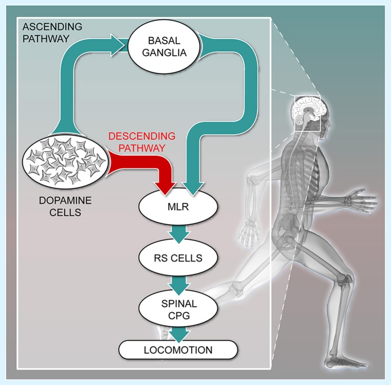
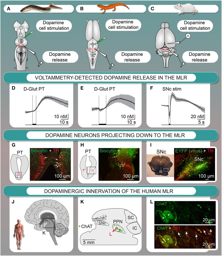
In vertebrates, dopamine neurons are classically known to modulate locomotion via their ascending projections to the basal ganglia that project to brainstem locomotor networks. An increased dopaminergic tone is associated with increase in locomotor activity. In pathological conditions where dopamine cells are lost, such as in Parkinson's disease, locomotor deficits are traditionally associated with the reduced ascending dopaminergic input to the basal ganglia. However, a descending dopaminergic pathway originating from the was recently discovered. It innervates the mesencephalic locomotor region (MLR) from basal vertebrates to mammals. This pathway was shown to increase locomotor output in lampreys, and could very well play an important role in mammals. Here, we provide a detailed account on the newly found dopaminergic pathway in lamprey, salamander, rat, monkey, and human. In lampreys and salamanders, dopamine release in the MLR is associated with the activation of reticulospinal neurons that carry the locomotor command to the spinal cord. Dopamine release in the MLR potentiates locomotor movements through a D1-receptor mechanism in lampreys. In rats, stimulation of the elicited dopamine release in the pedunculopontine nucleus, a known part of the MLR. In a monkey model of Parkinson's disease, a reduced dopaminergic innervation of the brainstem locomotor networks was reported. Dopaminergic fibers are also present in human pedunculopontine nucleus. We discuss the conserved locomotor role of this pathway from lamprey to mammals, and the hypothesis that this pathway could play a role in the locomotor deficits reported in Parkinson's disease.
在脊椎动物中,多巴胺能神经元传统上被认为是通过其向基底神经节的上行投射来调节运动,而基底神经节又投射到脑干运动网络。多巴胺能张力增加与运动活动增加相关。在多巴胺细胞缺失的病理状况下,如帕金森病,运动缺陷传统上被认为与基底神经节的上行多巴胺能输入减少有关。然而,最近发现了一条源自 的下行多巴胺能通路。它从低等脊椎动物到哺乳动物都支配中脑运动区(MLR)。这条通路已被证明能增加七鳃鳗的运动输出,并且很可能在哺乳动物中发挥重要作用。在这里,我们详细阐述了在七鳃鳗、蝾螈、大鼠、猴子和人类中 newly found dopaminergic pathway(新发现的多巴胺能通路)。在七鳃鳗和蝾螈中,MLR中的多巴胺释放与将运动指令传递到脊髓的网状脊髓神经元的激活有关。在七鳃鳗中,MLR中的多巴胺释放通过D1受体机制增强运动。在大鼠中,刺激 会在脚桥核(MLR的一个已知部分)引发多巴胺释放。在帕金森病的猴子模型中,有报道称脑干运动网络的多巴胺能神经支配减少。人类脚桥核中也存在多巴胺能纤维。我们讨论了这条从七鳃鳗到哺乳动物的通路在运动方面的保守作用,以及该通路可能在帕金森病所报道的运动缺陷中起作用的假说。