• 文献检索
  • 文档翻译
  • 深度研究
  • 学术资讯
  • Suppr Zotero 插件Zotero 插件
  • 邀请有礼
  • 套餐&价格
  • 历史记录
应用&插件
Suppr Zotero 插件Zotero 插件浏览器插件Mac 客户端Windows 客户端微信小程序
定价
高级版会员购买积分包购买API积分包
服务
文献检索文档翻译深度研究API 文档MCP 服务
关于我们
关于 Suppr公司介绍联系我们用户协议隐私条款
关注我们

Suppr 超能文献

核心技术专利:CN118964589B侵权必究
粤ICP备2023148730 号-1Suppr @ 2026

文献检索

告别复杂PubMed语法,用中文像聊天一样搜索,搜遍4000万医学文献。AI智能推荐,让科研检索更轻松。

立即免费搜索

文件翻译

保留排版,准确专业,支持PDF/Word/PPT等文件格式,支持 12+语言互译。

免费翻译文档

深度研究

AI帮你快速写综述,25分钟生成高质量综述,智能提取关键信息,辅助科研写作。

立即免费体验

使用对比增强微计算机断层扫描对骨软骨组织进行 3D 组织病理学分级。

3D histopathological grading of osteochondral tissue using contrast-enhanced micro-computed tomography.

机构信息

Research Unit of Medical Imaging, Physics and Technology, Faculty of Medicine, University of Oulu, Oulu, Finland; Department of Laboratory Medicine and Pathobiology, University of Toronto, Toronto, Canada; Department of Physics, University of Helsinki, Helsinki, Finland; Department of Neuroscience and Biomedical Engineering, Aalto University, Espoo, Finland.

Orthopedic Science Consulting Services, Oakville, Ontario, Canada.

出版信息

Osteoarthritis Cartilage. 2017 Oct;25(10):1680-1689. doi: 10.1016/j.joca.2017.05.021. Epub 2017 Jun 9.

DOI:10.1016/j.joca.2017.05.021
PMID:28606558
原文链接:https://pmc.ncbi.nlm.nih.gov/articles/PMC5773475/
Abstract

OBJECTIVE

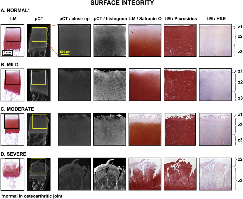
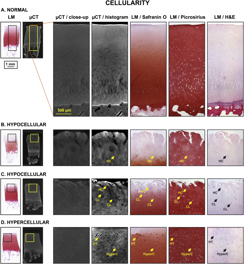
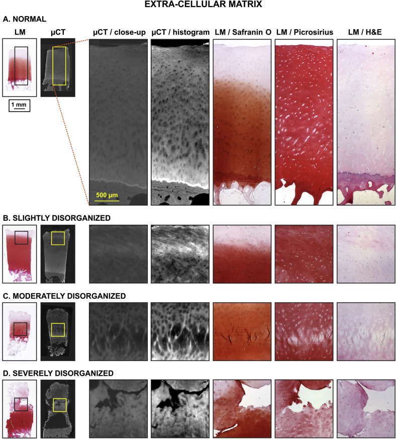
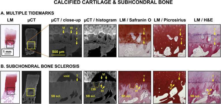
Histopathological grading of osteochondral (OC) tissue is widely used in osteoarthritis (OA) research, and it is relatively common in post-surgery in vitro diagnostics. However, relying on thin tissue section, this approach includes a number of limitations, such as: (1) destructiveness, (2) sample processing artefacts, (3) 2D section does not represent spatial 3D structure and composition of the tissue, and (4) the final outcome is subjective. To overcome these limitations, we recently developed a contrast-enhanced μCT (CEμCT) imaging technique to visualize the collagenous extracellular matrix (ECM) of articular cartilage (AC). In the present study, we demonstrate that histopathological scoring of OC tissue from CEμCT is feasible. Moreover, we establish a new, semi-quantitative OA μCT grading system for OC tissue.

RESULTS

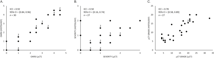
Pathological features were clearly visualized in AC and subchondral bone (SB) with μCT and verified with histology, as demonstrated with image atlases. Comparison of histopathological grades (OARSI or severity (0-3)) across the characterization approaches, CEμCT and histology, excellent (0.92, 95% CI = [0.84, 0.96], n = 30) or fair (0.50, 95% CI = [0.16, 0.74], n = 27) intra-class correlations (ICC), respectively. A new μCT grading system was successfully established which achieved an excellent cross-method (μCT vs histology) reader-to-reader intra-class correlation (0.78, 95% CI = [0.58, 0.89], n = 27).

CONCLUSIONS

We demonstrated that histopathological information relevant to OA can reliably be obtained from CEμCT images. This new grading system could be used as a reference for 3D imaging and analysis techniques intended for volumetric evaluation of OA pathology in research and clinical applications.

摘要

目的

骨软骨(OC)组织的组织病理学分级在骨关节炎(OA)研究中被广泛应用,并且在术后体外诊断中也较为常见。然而,这种方法依赖于薄的组织切片,存在许多局限性,例如:(1)破坏性,(2)样本处理伪影,(3)2D 切片不能代表组织的空间 3D 结构和组成,以及(4)最终结果是主观的。为了克服这些局限性,我们最近开发了一种对比增强 μCT(CEμCT)成像技术来可视化关节软骨(AC)的胶原细胞外基质(ECM)。在本研究中,我们证明了从 CEμCT 对 OC 组织进行组织病理学评分是可行的。此外,我们建立了一种新的、半定量的 OC 组织 OA μCT 分级系统。

结果

通过 μCT 和组织学验证,使用图像图谱清楚地观察到 AC 和软骨下骨(SB)的病理特征,与组织学一致。在组织学和 CEμCT 两种方法中,OC 组织的组织病理学分级(OARSI 或严重程度(0-3))之间具有极好(0.92,95%CI[0.84,0.96],n=30)或良好(0.50,95%CI[0.16,0.74],n=27)的一致性(ICC)。成功建立了新的 μCT 分级系统,其在跨方法(μCT 与组织学)读者间的一致性极好(0.78,95%CI[0.58,0.89],n=27)。

结论

我们证明了与 OA 相关的组织病理学信息可以从 CEμCT 图像中可靠地获得。这种新的分级系统可作为用于 OA 病理学体积评估的 3D 成像和分析技术的参考。

https://cdn.ncbi.nlm.nih.gov/pmc/blobs/b727/5773475/710e5cd3dcb7/gr5.jpg
https://cdn.ncbi.nlm.nih.gov/pmc/blobs/b727/5773475/9510096b77a2/gr1.jpg
https://cdn.ncbi.nlm.nih.gov/pmc/blobs/b727/5773475/bddd053dc46d/gr2.jpg
https://cdn.ncbi.nlm.nih.gov/pmc/blobs/b727/5773475/82ac32769d6a/gr3.jpg
https://cdn.ncbi.nlm.nih.gov/pmc/blobs/b727/5773475/aea007cbf6de/gr4.jpg
https://cdn.ncbi.nlm.nih.gov/pmc/blobs/b727/5773475/710e5cd3dcb7/gr5.jpg

相似文献

1
3D histopathological grading of osteochondral tissue using contrast-enhanced micro-computed tomography.使用对比增强微计算机断层扫描对骨软骨组织进行 3D 组织病理学分级。
Osteoarthritis Cartilage. 2017 Oct;25(10):1680-1689. doi: 10.1016/j.joca.2017.05.021. Epub 2017 Jun 9.
2
Automating three-dimensional osteoarthritis histopathological grading of human osteochondral tissue using machine learning on contrast-enhanced micro-computed tomography.利用对比增强微计算机断层扫描的机器学习,实现人类骨软骨组织三维骨关节炎组织病理学分级的自动化。
Osteoarthritis Cartilage. 2020 Aug;28(8):1133-1144. doi: 10.1016/j.joca.2020.05.002. Epub 2020 May 11.
3
3D morphometric analysis of calcified cartilage properties using micro-computed tomography.使用微计算机断层扫描对钙化软骨特性进行 3D 形态计量分析。
Osteoarthritis Cartilage. 2019 Jan;27(1):172-180. doi: 10.1016/j.joca.2018.09.009. Epub 2018 Oct 1.
4
Association between subchondral bone structure and osteoarthritis histopathological grade.软骨下骨结构与骨关节炎组织病理学分级之间的关联。
J Orthop Res. 2017 Apr;35(4):785-792. doi: 10.1002/jor.23312. Epub 2016 Jun 22.
5
3D analysis and grading of calcifications from ex vivo human meniscus.离体人半月板钙化的 3D 分析和分级。
Osteoarthritis Cartilage. 2023 Apr;31(4):482-492. doi: 10.1016/j.joca.2022.10.016. Epub 2022 Nov 7.
6
Quantifying Complex Micro-Topography of Degenerated Articular Cartilage Surface by Contrast-Enhanced Micro-Computed Tomography and Parametric Analyses.采用对比增强微计算机断层扫描和参数分析定量评估退变关节软骨表面的复杂微观形貌。
J Orthop Res. 2019 Apr;37(4):855-866. doi: 10.1002/jor.24245. Epub 2019 Mar 5.
7
Varying development of femoral and tibial subchondral bone tissue and their interaction with articular cartilage during progressing osteoarthritis.在进展性骨关节炎中,股骨和胫骨的软骨下骨组织的不同发育及其与关节软骨的相互作用。
Arch Orthop Trauma Surg. 2020 Dec;140(12):1919-1930. doi: 10.1007/s00402-020-03480-w. Epub 2020 May 30.
8
Bone turnover and articular cartilage differences localized to subchondral cysts in knees with advanced osteoarthritis.骨转换和关节软骨差异定位于晚期骨关节炎膝关节的软骨下囊肿。
Osteoarthritis Cartilage. 2015 Dec;23(12):2174-2183. doi: 10.1016/j.joca.2015.07.012. Epub 2015 Aug 1.
9
In vitro method for 3D morphometry of human articular cartilage chondrons based on micro-computed tomography.基于微计算机断层扫描的人关节软骨软骨细胞 3D 形态计量学的体外方法。
Osteoarthritis Cartilage. 2018 Aug;26(8):1118-1126. doi: 10.1016/j.joca.2018.05.012. Epub 2018 May 23.
10
Subchondral bone scan uptake correlates with articular cartilage degeneration in osteoarthritic knees.骨关节炎膝关节的软骨下骨扫描摄取与关节软骨退变相关。
Int J Rheum Dis. 2017 Oct;20(10):1393-1402. doi: 10.1111/1756-185X.12909. Epub 2016 Jul 25.

引用本文的文献

1
Contrast-enhanced Micro-CT 3D visualization of cell distribution in hydrated human cornea.水合人角膜中细胞分布的对比增强微型计算机断层扫描三维可视化
Heliyon. 2024 Feb 3;10(3):e25828. doi: 10.1016/j.heliyon.2024.e25828. eCollection 2024 Feb 15.
2
Optimal Agents for Visualizing Collagen Tissue Microarchitecture Using Contrast-Enhanced MicroCT.使用对比增强显微CT可视化胶原组织微观结构的最佳试剂
Pharmaceuticals (Basel). 2023 Dec 12;16(12):1719. doi: 10.3390/ph16121719.
3
Influence of fixation on CA4+ contrast enhanced microCT of articular cartilage and subsequent feasibility for histological evaluation.

本文引用的文献

1
Effects of Articular Cartilage Constituents on Phosphotungstic Acid Enhanced Micro-Computed Tomography.关节软骨成分对磷钨酸增强型微型计算机断层扫描的影响。
PLoS One. 2017 Jan 30;12(1):e0171075. doi: 10.1371/journal.pone.0171075. eCollection 2017.
2
Systematic mapping of the subchondral bone 3D microarchitecture in the human tibial plateau: Variations with joint alignment.人胫骨平台软骨下骨三维微结构的系统映射:随关节对线的变化
J Orthop Res. 2017 Sep;35(9):1927-1941. doi: 10.1002/jor.23474. Epub 2016 Nov 28.
3
Association between subchondral bone structure and osteoarthritis histopathological grade.
固定对关节软骨CA4 +对比增强微计算机断层扫描的影响及后续组织学评估的可行性。
Am J Transl Res. 2021 Aug 15;13(8):8921-8937. eCollection 2021.
4
Contrast-Enhanced MicroCT for Virtual 3D Anatomical Pathology of Biological Tissues: A Literature Review.对比增强微 CT 在生物组织虚拟 3D 解剖病理学中的应用:文献综述。
Contrast Media Mol Imaging. 2019 Feb 28;2019:8617406. doi: 10.1155/2019/8617406. eCollection 2019.
5
Quantifying Complex Micro-Topography of Degenerated Articular Cartilage Surface by Contrast-Enhanced Micro-Computed Tomography and Parametric Analyses.采用对比增强微计算机断层扫描和参数分析定量评估退变关节软骨表面的复杂微观形貌。
J Orthop Res. 2019 Apr;37(4):855-866. doi: 10.1002/jor.24245. Epub 2019 Mar 5.
6
3D vessel-wall virtual histology of whole-body perfused mice using a novel heavy element stain.使用新型重元素染色剂对全身灌注小鼠进行 3D 血管壁虚拟组织学研究。
Sci Rep. 2019 Jan 24;9(1):698. doi: 10.1038/s41598-018-36905-z.
7
In vitro method for 3D morphometry of human articular cartilage chondrons based on micro-computed tomography.基于微计算机断层扫描的人关节软骨软骨细胞 3D 形态计量学的体外方法。
Osteoarthritis Cartilage. 2018 Aug;26(8):1118-1126. doi: 10.1016/j.joca.2018.05.012. Epub 2018 May 23.
软骨下骨结构与骨关节炎组织病理学分级之间的关联。
J Orthop Res. 2017 Apr;35(4):785-792. doi: 10.1002/jor.23312. Epub 2016 Jun 22.
4
3D ultrasound biomicroscopy for assessment of cartilage repair tissue: volumetric characterisation and correlation to established classification systems.用于评估软骨修复组织的三维超声生物显微镜检查:体积特征及与既定分类系统的相关性
Eur Cell Mater. 2016 Feb 8;31:119-35. doi: 10.22203/ecm.v031a09.
5
Depth-Dependent Glycosaminoglycan Concentration in Articular Cartilage by Quantitative Contrast-Enhanced Micro-Computed Tomography.通过定量对比增强微型计算机断层扫描术测定关节软骨中糖胺聚糖浓度的深度依赖性
Cartilage. 2015 Oct;6(4):216-25. doi: 10.1177/1947603515596418.
6
Correcting the Shrinkage Effects of Formalin Fixation and Tissue Processing for Renal Tumors: toward Standardization of Pathological Reporting of Tumor Size.纠正福尔马林固定和组织处理对肾肿瘤的收缩效应:实现肿瘤大小病理报告标准化。
J Cancer. 2015 Jul 2;6(8):759-66. doi: 10.7150/jca.12094. eCollection 2015.
7
Determining collagen distribution in articular cartilage using contrast-enhanced micro-computed tomography.使用对比增强型微型计算机断层扫描确定关节软骨中的胶原蛋白分布。
Osteoarthritis Cartilage. 2015 Sep;23(9):1613-21. doi: 10.1016/j.joca.2015.05.004. Epub 2015 May 21.
8
How best to preserve and reveal the structural intricacies of cartilaginous tissue.如何最好地保存和揭示软骨组织的结构复杂性。
Matrix Biol. 2014 Oct;39:33-43. doi: 10.1016/j.matbio.2014.08.010. Epub 2014 Aug 27.
9
In vivo diagnostics of human knee cartilage lesions using delayed CBCT arthrography.利用延迟 CBCT 关节造影术对人体膝关节软骨病变进行体内诊断。
J Orthop Res. 2014 Mar;32(3):403-12. doi: 10.1002/jor.22521. Epub 2013 Nov 19.
10
Reconstruction of three dimensional structures from electron micrographs.从电子显微镜照片重建三维结构。
Nature. 1968 Jan 13;217(5124):130-4. doi: 10.1038/217130a0.