Research Unit of Medical Imaging, Physics and Technology, Faculty of Medicine, University of Oulu, Oulu, Finland.
Department of Physics, University of Helsinki, Helsinki, Finland.
J Orthop Res. 2019 Apr;37(4):855-866. doi: 10.1002/jor.24245. Epub 2019 Mar 5.
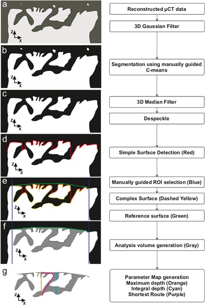
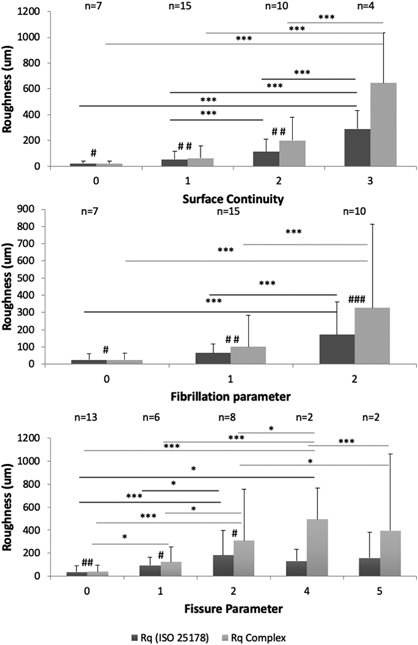
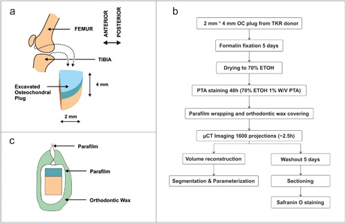
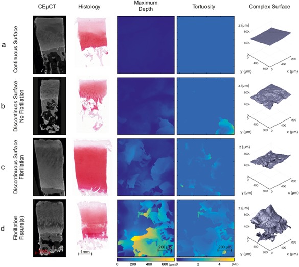
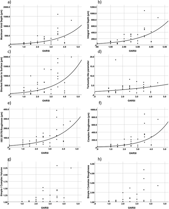
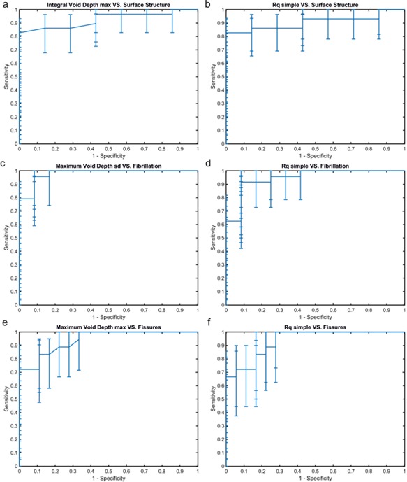
One of the earliest changes in osteoarthritis (OA) is a surface discontinuity of the articular cartilage (AC), and these surface changes become gradually more complex with OA progression. We recently developed a contrast enhanced micro-computed tomography (μCT) method for visualizing AC surface in detail. The present study aims to introduce a μCT analysis technique to parameterize these complex AC surface features and to demonstrate the feasibility of using these parameters to quantify degenerated AC surface. Osteochondral plugs (n = 35) extracted from 19 patients undergoing joint surgery were stained with phosphotungstic acid and imaged using μCT. The surface micro-topography of AC was analyzed with developed method. Standard root mean square roughness (R ) was calculated as a reference, and the Area Under Curve (AUC) for receiver operating characteristic analysis was used to compare the acquired quantitative parameters with semi-quantitative visual grading of μCT image stacks. The parameters quantifying the complex micro-topography of AC surface exhibited good sensitivity and specificity in identifying surface continuity (AUC: 0.93, [0.80 0.99]), fissures (AUC: 0.94, [0.83 0.99]) and fibrillation (AUC: 0.98, [0.88 1.0]). Standard R was significantly smaller compared with the complex roughness (CR ) already with mild surface changes with all surface reference parameters - continuity, fibrillation, and fissure sum. Furthermore, only CR showed a significant difference when comparing the intact surface with lowest fissure sum score. These results indicate that the presented method for evaluating complex AC surfaces exhibit potential to identify early OA changes in superficial AC and is dynamic throughout OA progression. © 2019 The Authors. Journal of Orthopaedic Research® Published by Wiley Periodicals, Inc. on behalf of the Orthopaedic Research Society. Society. 9999:1-12, 2019.
骨关节炎(OA)最早的变化之一是关节软骨(AC)的表面不连续性,并且这些表面变化随着 OA 的进展而变得越来越复杂。我们最近开发了一种对比度增强的微计算机断层扫描(μCT)方法来详细观察 AC 表面。本研究旨在介绍一种μCT 分析技术来参数化这些复杂的 AC 表面特征,并证明使用这些参数来量化退变 AC 表面的可行性。从接受关节手术的 19 名患者中提取的骨软骨嵌块(n=35)用磷钨酸染色,并使用μCT 成像。使用开发的方法分析 AC 的表面微观形貌。计算了标准均方根粗糙度(R)作为参考,并用接收器操作特性分析的曲线下面积(AUC)来比较获得的定量参数与μCT 图像堆栈的半定量视觉分级。定量 AC 表面复杂微观形貌的参数在识别表面连续性(AUC:0.93 [0.80-0.99])、裂隙(AUC:0.94 [0.83-0.99])和纤维化(AUC:0.98 [0.88-1.0])方面表现出良好的灵敏度和特异性。标准 R 与轻度表面变化时的复杂粗糙度(CR)相比已经明显减小,所有表面参考参数 - 连续性、纤维化和裂隙总和。此外,当比较具有最低裂隙总和评分的完整表面时,只有 CR 显示出显著差异。这些结果表明,评估复杂 AC 表面的方法具有识别浅层 AC 早期 OA 变化的潜力,并在 OA 进展过程中具有动态性。 2019 年,作者。《矫形研究杂志》由 Wiley 期刊出版公司代表矫形研究协会出版。9999:1-12,2019。