La Marra Francesco, Stinco Giuseppe, Buligan Cinzia, Chiriacò Giovanni, Serraino Diego, Di Loreto Carla, Cauci Sabina
Department of Medical Area, School of Medicine, University of Udine, Udine 33100, Italy.
Dermatology Clinic, Udine University-Hospital, University of Udine, Udine 33100, Italy.
Cancer Biol Med. 2017 May;14(2):162-175. doi: 10.20892/j.issn.2095-3941.2017.0020.
: Vitamin D receptor (VDR) mediates vitamin D activity. We examined whether VDR expression in excised melanoma tissues is associated with VDR gene () polymorphisms.
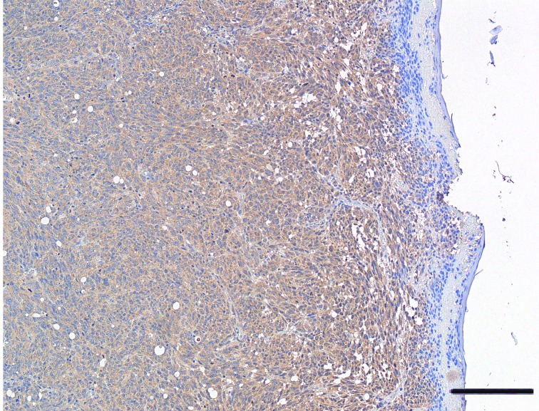
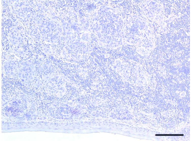
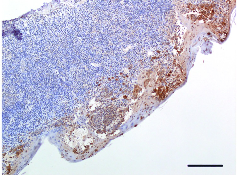
: We evaluated VDR protein expression (by monoclonal antibody immunostaining), melanoma characteristics, and carriage of -FokI-rs2228570 (C>T),-BsmI-rs1544410 (G>A),-ApaI-rs7975232 (T>G), and-TaqI-rs731236 (T>C) polymorphisms (by restriction fragment length polymorphism). Absence or presence of restriction site was denoted by a capital or lower letter, respectively: " F" and " f" for FokI, " B" and " b" for BsmI, " A" and " a" for ApaI, and " T" and " t" for TaqI endonuclease. Seventy-four Italian cutaneous primary melanomas (52.1±12.7 years old) were studied; 51.4% were stage I, 21.6% stage II, 13.5% stage III, and 13.5% stage IV melanomas. VDR expression was categorized as follows: 100% positive. <100%; over the median 20% (high VDR expression) . ≤20% (low VDR expression); absence . presence of VDR-expressing cells.
: Stage I melanomas, Breslow thickness of <1.00 mm, level II Clark invasion, Aa heterozygous genotype, and AaTT combined genotype were more frequent in melanomas with high . low VDR expression. Combined genotypes BbAA, bbAa, AATt, BbAATt, and bbAaTT were more frequent in 100% . <100% VDR-expressing cells. Combined genotype AATT was more frequent in melanomas lacking VDR expression (odds ratio=14.5; =0.025). VDR expression was not associated with metastasis, ulceration, mitosis >1, regression, tumor-infiltrating lymphocytes, tumoral infiltration of vascular tissues, additional skin and non-skin cancers, and melanoma familiarity.
: We highlighted that polymorphisms can affect VDR expression in excised melanoma cells. Low VDR expression in AATT carriers is a new finding that merits further study. VDR expression possibly poses implications for vitamin D supplementation against melanoma. VDR expression and genotype may become precise medicinal tools for melanoma in the future.
维生素D受体(VDR)介导维生素D的活性。我们研究了切除的黑色素瘤组织中VDR的表达是否与VDR基因()多态性相关。
我们评估了VDR蛋白表达(通过单克隆抗体免疫染色)、黑色素瘤特征以及-FokI-rs2228570(C>T)、-BsmI-rs1544410(G>A)、-ApaI-rs7975232(T>G)和-TaqI-rs731236(T>C)多态性(通过限制性片段长度多态性)。限制性位点的缺失或存在分别用大写或小写字母表示:FokI的“F”和“f”、BsmI的“B”和“b”、ApaI的“A”和“a”以及TaqI核酸内切酶的“T”和“t”。研究了74例意大利皮肤原发性黑色素瘤(年龄52.1±12.7岁);51.4%为I期,21.6%为II期,13.5%为III期,13.5%为IV期黑色素瘤。VDR表达分类如下:100%阳性;<100%;超过中位数20%(高VDR表达);≤20%(低VDR表达);无VDR表达细胞;有VDR表达细胞。
I期黑色素瘤、Breslow厚度<1.00mm、Clark II级浸润、Aa杂合基因型以及AaTT联合基因型在高VDR表达、低VDR表达的黑色素瘤中更常见。联合基因型BbAA、bbAa、AATt、BbAATt和bbAaTT在VDR表达细胞为100%、<100%时更常见。联合基因型AATT在缺乏VDR表达的黑色素瘤中更常见(优势比=14.5;P=0.025)。VDR表达与转移、溃疡、有丝分裂>1、消退、肿瘤浸润淋巴细胞、血管组织的肿瘤浸润、其他皮肤和非皮肤癌以及黑色素瘤家族史无关。
我们强调基因多态性可影响切除的黑色素瘤细胞中VDR的表达。AATT携带者中VDR低表达是一个值得进一步研究的新发现。VDR表达可能对补充维生素D预防黑色素瘤有影响。VDR表达和基因型未来可能成为黑色素瘤精确的医学工具。