Linderman Rachel, Salmon Alexander E, Strampe Margaret, Russillo Madia, Khan Jamil, Carroll Joseph
Department of Ophthalmology & Visual Sciences, Medical College of Wisconsin, Milwaukee, WI, USA.
Department of Cell Biology, Neurobiology, & Anatomy, Medical College of Wisconsin, Milwaukee, WI, USA.
Transl Vis Sci Technol. 2017 Jun 9;6(3):16. doi: 10.1167/tvst.6.3.16. eCollection 2017 Jun.
The foveal avascular zone (FAZ) is altered in numerous diseases. We assessed factors (axial length, segmentation method, age, sex) impacting FAZ measurements from optical coherence tomography (OCT) angiography images.
We recruited 116 Caucasian subjects without ocular disease, and acquired two 3 × 3 mm AngioVue scans per each right eye (232 total scans). In images of the superficial plexus, the FAZ was segmented using the AngioVue semiautomatic nonflow measurement tool and ImageJ manual segmentation. In images from the full retinal thickness, the FAZ was segmented using the AngioAnalytics automatic FAZ tool. Repeatability, reliability, and reproducibility were calculated for FAZ measurements (acircularity, area).
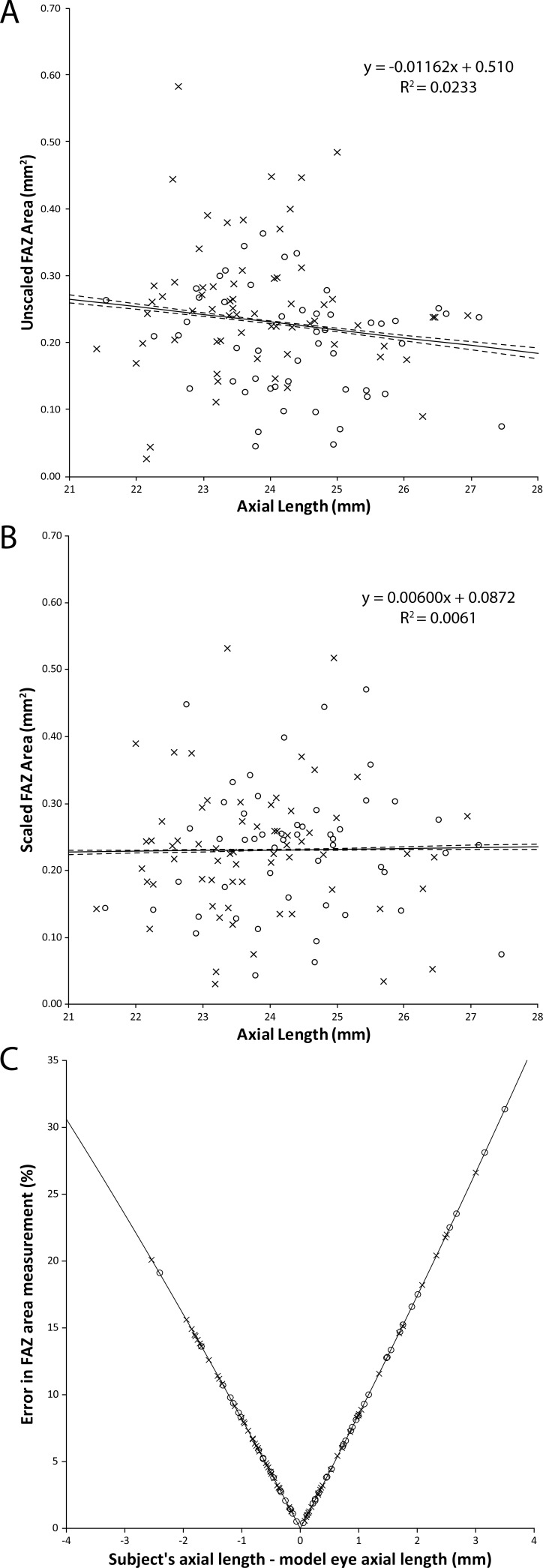
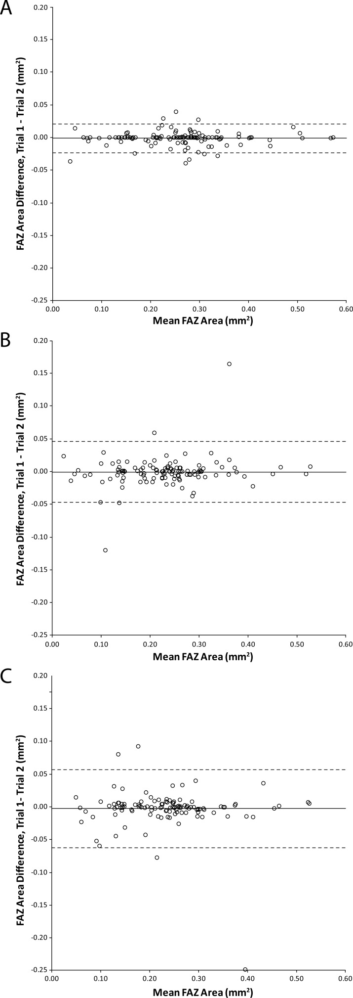
FAZ area (mean ± SD) for manual segmentation was 0.240 ± 0.0965 mm, greater than both semiautomatic (0.216 ± 0.0873 mm) and automatic (0.218 ± 0.0869 mm) segmentation ( < 0.05). Not correcting for axial length introduced errors up to 25% in FAZ area. Manual area segmentation had better repeatability (0.020 mm) than semiautomatic (0.043 mm) or automatic (0.056 mm). FAZ acircularity had better repeatability with automatic than manual segmentation (0.086 vs. 0.114). Reliability of all area measurements was excellent (intraclass correlation coefficient [ICC] = 0.994 manual, 0.969 semiautomatic, 0.948 automatic). Reliability of acircularity measurements was 0.879 for manual and 0.606 for automatic.
We identified numerous factors affecting FAZ measurements. These errors confound comparisons across studies and studies examining factors that may correlate with FAZ measures.
Using FAZ measurements as biomarkers for disease progression requires assessing and controlling for different sources of error. Not correcting for ocular magnification can result in significant inaccuracy in FAZ measurements, while choice of segmentation method affects both repeatability and accuracy.
黄斑无血管区(FAZ)在多种疾病中会发生改变。我们评估了影响光学相干断层扫描血管造影(OCTA)图像中FAZ测量的因素(眼轴长度、分割方法、年龄、性别)。
我们招募了116名无眼部疾病的白种人受试者,每只右眼获取两次3×3mm的AngioVue扫描图像(共232次扫描)。在浅层血管丛图像中,使用AngioVue半自动非血流测量工具和ImageJ手动分割法对FAZ进行分割。在全视网膜厚度图像中,使用AngioAnalytics自动FAZ工具对FAZ进行分割。计算FAZ测量值(非圆度、面积)的重复性、可靠性和再现性。
手动分割的FAZ面积(均值±标准差)为0.240±0.0965mm²,大于半自动分割(0.216±0.0873mm²)和自动分割(0.218±0.0869mm²)(P<0.05)。未校正眼轴长度会导致FAZ面积测量误差高达25%。手动面积分割的重复性(0.020mm²)优于半自动分割(0.043mm²)和自动分割(0.056mm²)。FAZ非圆度自动分割的重复性优于手动分割(0.086对0.114)。所有面积测量的可靠性都非常好(组内相关系数[ICC]:手动分割为0.994,半自动分割为0.969,自动分割为0.948)。非圆度测量的可靠性,手动分割为0.879,自动分割为0.606。
我们确定了影响FAZ测量的多种因素。这些误差混淆了不同研究之间以及研究中检查可能与FAZ测量相关因素的比较。
将FAZ测量用作疾病进展的生物标志物需要评估和控制不同的误差来源。未校正眼放大率会导致FAZ测量出现显著误差,而分割方法的选择会影响重复性和准确性。