Lapinskas Tomas, Schnackenburg Bernhard, Kouwenhoven Marc, Gebker Rolf, Berger Alexander, Zaliunas Remigijus, Pieske Burkert, Kelle Sebastian
Department of Cardiology, Medical Academy, Lithuanian University of Health Sciences, Eiveniu Street 2, 50161, Kaunas, Lithuania.
Department of Internal Medicine/Cardiology, German Heart Institute Berlin, Augustenburger Platz 1, 13353, Berlin, Germany.
MAGMA. 2018 Feb;31(1):75-85. doi: 10.1007/s10334-017-0639-7. Epub 2017 Jun 15.
This study aimed to investigate the advantages of recently developed cardiac imaging techniques of fat-water separation and feature tracking to characterize better individuals with chronic myocardial infarction (MI).
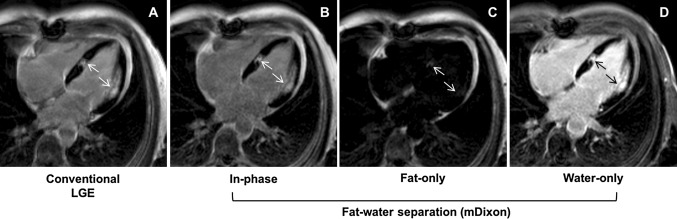
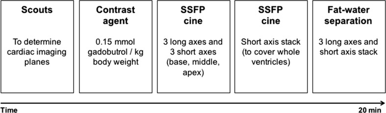
Twenty patients who had a previous MI underwent CMR imaging. The study protocol included routine cine and late gadolinium enhancement (LGE) technique. In addition, mDixon LGE imaging was performed in every patient. Left ventricular (LV) circumferential (Ecc) and radial (Err) strain were calculated using dedicated software (CMR, Circle, Calgary, Canada). The extent of global scar was measured in LGE and fat-water separated images to compare conventional and recent CMR imaging techniques.
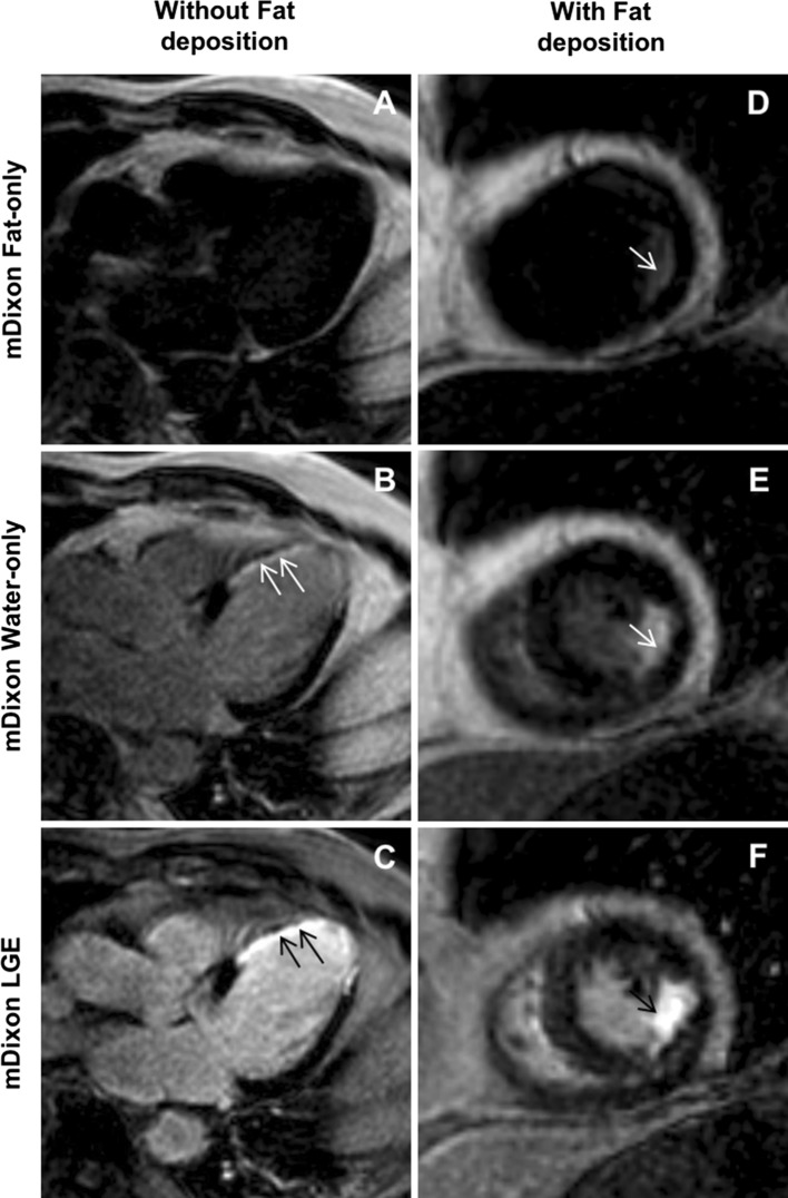
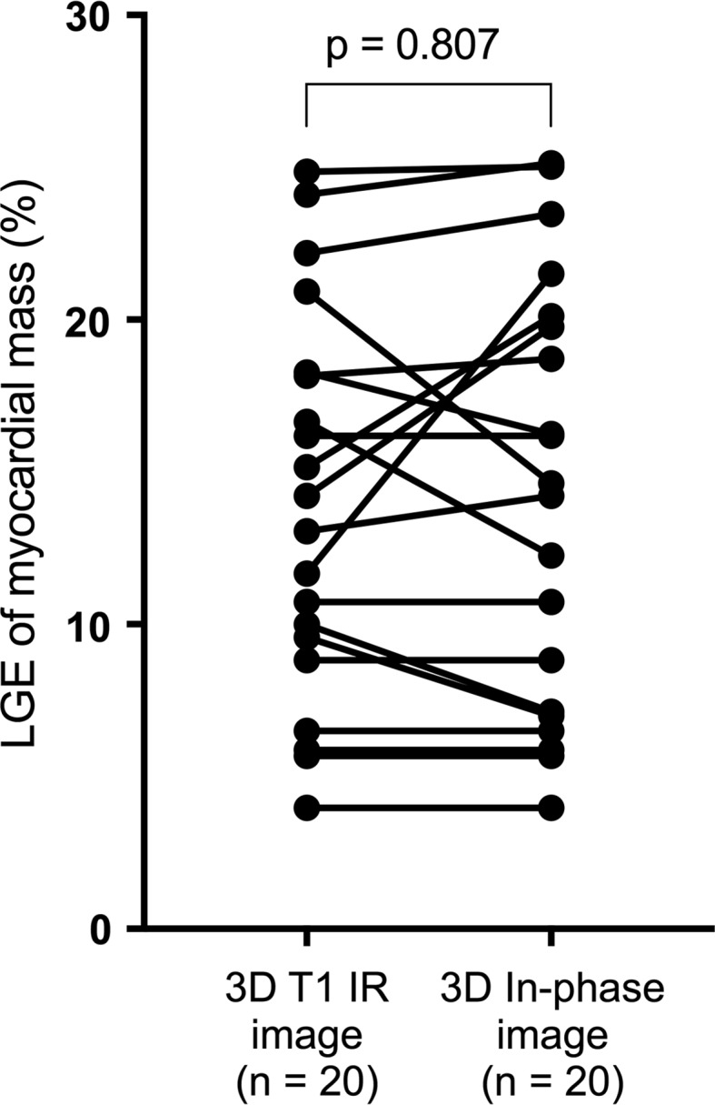
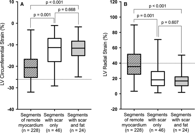
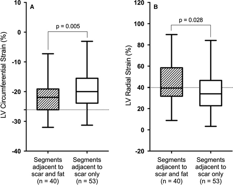
The infarct size derived from conventional LGE and fat-water separated images was similar. However, detection of lipomatous metaplasia was only possible with mDixon imaging. Subjects with fat deposition demonstrated a significantly smaller percentage of fibrosis than those without fat (10.68 ± 5.07% vs. 13.83 ± 6.30%; p = 0.005). There was no significant difference in Ecc or Err between myocardial segments containing fibrosis only and fibrosis with fat. However, Ecc and Err values were significantly higher in myocardial segments adjacent to fibrosis with fat deposition than in those adjacent to LGE only.
Advanced CMR imaging ensures more detailed tissue characterization in patients with chronic MI without a relevant increase in imaging and post-processing time. Fatty metaplasia may influence regional myocardial deformation especially in the myocardial segments adjacent to scar tissue. A simplified and shortened myocardial viability CMR protocol might be useful to better characterize and stratify patients with chronic MI.
本研究旨在探讨近期开发的脂肪-水分离和特征追踪心脏成像技术在更好地表征慢性心肌梗死(MI)患者方面的优势。
20例曾患心肌梗死的患者接受了心脏磁共振成像(CMR)检查。研究方案包括常规电影成像和延迟钆增强(LGE)技术。此外,每位患者均进行了mDixon LGE成像。使用专用软件(CMR,Circle,加拿大卡尔加里)计算左心室(LV)圆周应变(Ecc)和径向应变(Err)。在LGE图像和脂肪-水分离图像中测量整体瘢痕范围,以比较传统CMR成像技术和近期CMR成像技术。
传统LGE图像和脂肪-水分离图像得出的梗死面积相似。然而,只有mDixon成像能够检测到脂肪化生。有脂肪沉积的受试者纤维化百分比明显低于无脂肪者(10.68±5.07%对13.83±6.30%;p = 0.005)。仅含纤维化的心肌节段与含脂肪纤维化的心肌节段之间的Ecc或Err无显著差异。然而,与仅LGE相邻的心肌节段相比,脂肪沉积纤维化相邻心肌节段的Ecc和Err值明显更高。
先进的CMR成像可确保在慢性心肌梗死患者中更详细地表征组织,而不会显著增加成像和后处理时间。脂肪化生可能影响局部心肌变形,尤其是在瘢痕组织相邻的心肌节段。简化和缩短的心肌存活CMR方案可能有助于更好地表征和分层慢性心肌梗死患者。