Research Center for Sectional and Imaging Anatomy, Shandong University School of Medicine, Jinan, China.
Radiology Research, Children's Hospital of Philadelphia, 3401 Civic Center Blvd, Philadelphia, PA, 19104, USA.
Brain Struct Funct. 2017 Dec;222(9):4131-4147. doi: 10.1007/s00429-017-1463-6. Epub 2017 Jun 20.
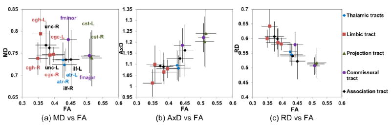
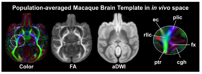
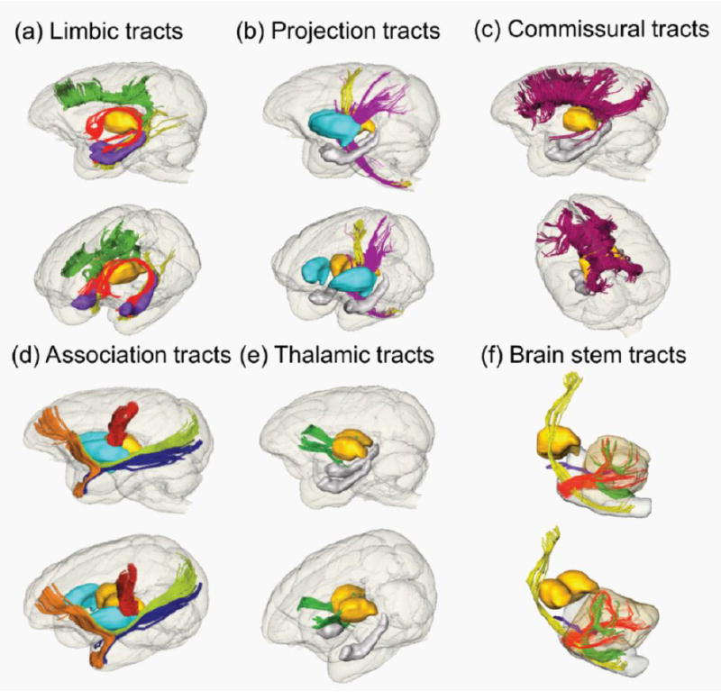
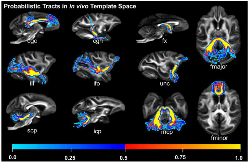
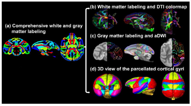
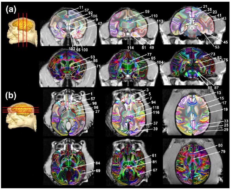
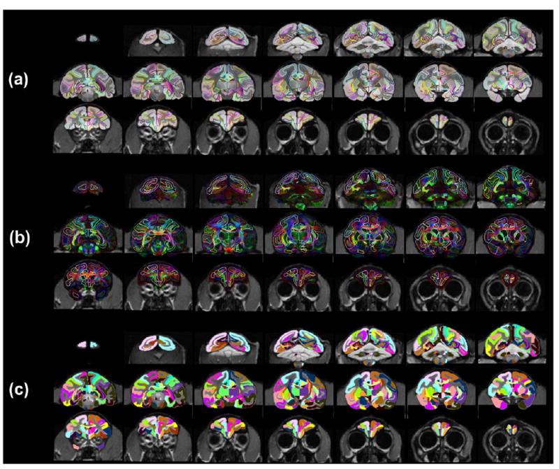
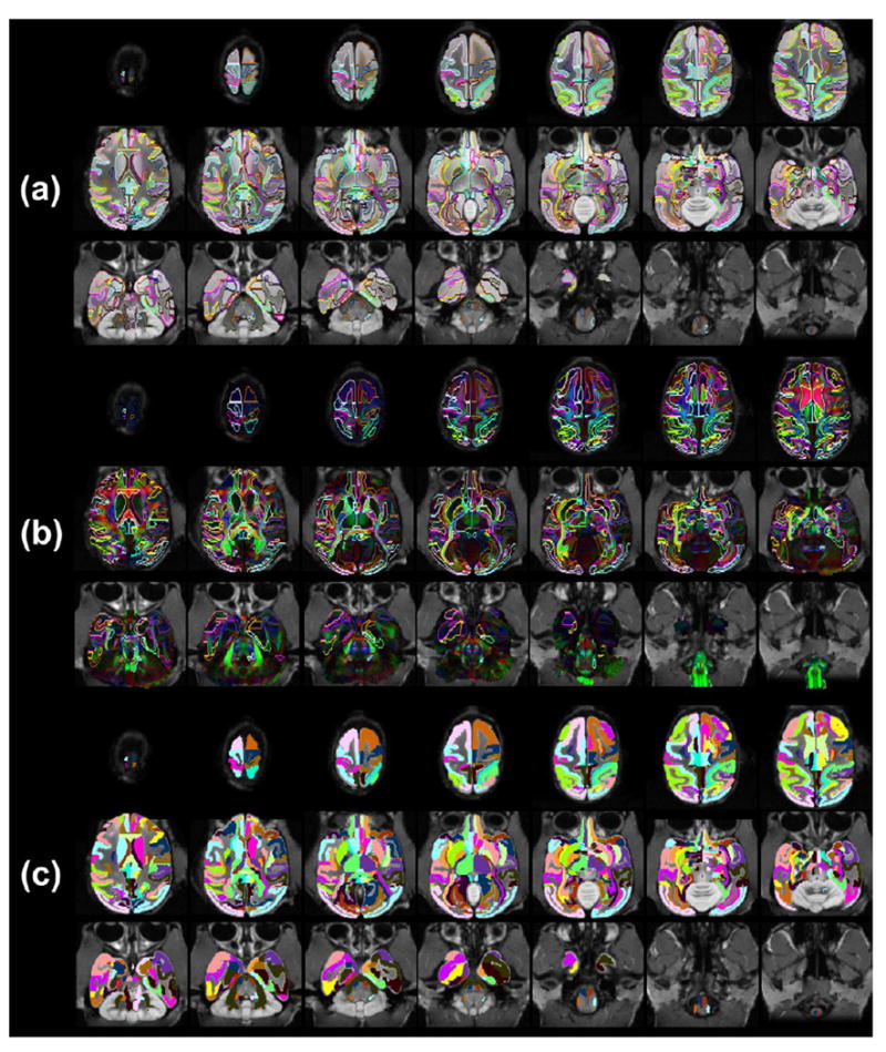
Animal models of the rhesus macaque (Macaca mulatta), the most widely used nonhuman primate, have been irreplaceable in neurobiological studies. However, a population-averaged macaque brain diffusion tensor imaging (DTI) atlas, including comprehensive gray and white matter labeling as well as bony and facial landmarks guiding invasive experimental procedures, is not available. The macaque white matter tract pathways and microstructures have been rarely recorded. Here, we established a population-averaged macaque brain atlas with high-resolution ex vivo DTI integrated into in vivo space incorporating bony and facial landmarks, and delineated microstructures and three-dimensional pathways of major white matter tracts in vivo MRI/DTI and ex vivo (postmortem) DTI of ten rhesus macaque brains were acquired. Single-subject macaque brain DTI template was obtained by transforming the postmortem high-resolution DTI data into in vivo space. Ex vivo DTI of ten macaque brains was then averaged in the in vivo single-subject template space to generate population-averaged macaque brain DTI atlas. The white matter tracts were traced with DTI-based tractography. One hundred and eighteen neural structures including all cortical gyri, white matter tracts and subcortical nuclei, were labeled manually on population-averaged DTI-derived maps. The in vivo microstructural metrics of fractional anisotropy, axial, radial and mean diffusivity of the traced white matter tracts were measured. Population-averaged digital atlas integrated into in vivo space can be used to label the experimental macaque brain automatically. Bony and facial landmarks will be available for guiding invasive procedures. The DTI metric measurements offer unique insights into heterogeneous microstructural profiles of different white matter tracts.
恒河猴(Macaca mulatta)作为最广泛使用的非人类灵长类动物,其动物模型在神经生物学研究中具有不可替代的作用。然而,目前尚缺乏包括全面的灰质和白质标记以及引导侵入性实验程序的骨性和面部标志的恒河猴大脑弥散张量成像(DTI)图谱。恒河猴的白质束通路和微观结构很少被记录下来。在这里,我们建立了一个基于群体的恒河猴大脑图谱,该图谱结合了骨性和面部标志,将高分辨率离体 DTI 与体内空间整合在一起,并对 10 只恒河猴大脑的体内 MRI/DTI 和离体(死后)DTI 进行了主要白质束的微观结构和三维通路的描绘。通过将死后高分辨率 DTI 数据转换到体内空间,获得了单只恒河猴大脑 DTI 模板。然后,在体内单只模板空间中对 10 只恒河猴大脑的离体 DTI 进行平均处理,生成基于群体的恒河猴大脑 DTI 图谱。使用基于 DTI 的束追踪技术对白质束进行追踪。在基于群体的 DTI 衍生图谱上手动标记了 118 个神经结构,包括所有皮质回、白质束和皮质下核。测量了追踪白质束的各向异性分数、轴向、径向和平均扩散率等微观结构指标。整合到体内空间的基于群体的数字图谱可用于自动标记实验恒河猴大脑。骨性和面部标志可用于引导侵入性程序。DTI 指标测量提供了不同白质束异质微观结构特征的独特见解。