Richard D. Berlin Center for Cell Analysis and Modeling, University of Connecticut School of Medicine, 400 Farmington Avenue, Farmington, Connecticut 06030, USA.
New England Musculoskeletal Institute, University of Connecticut School of Medicine, Farmington, Connecticut 06030, USA.
Nat Commun. 2017 Jun 21;8:15831. doi: 10.1038/ncomms15831.
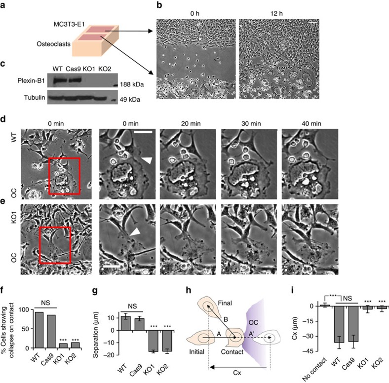
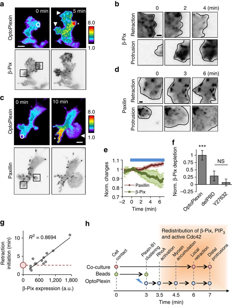
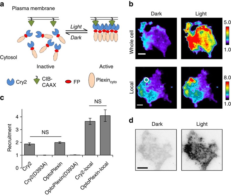
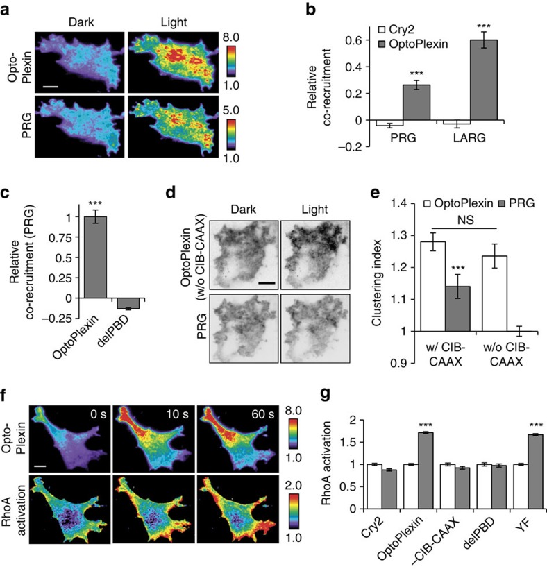
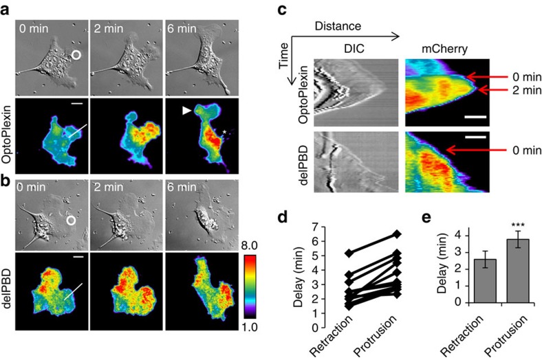
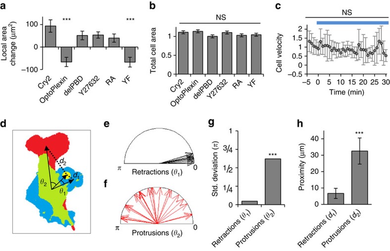
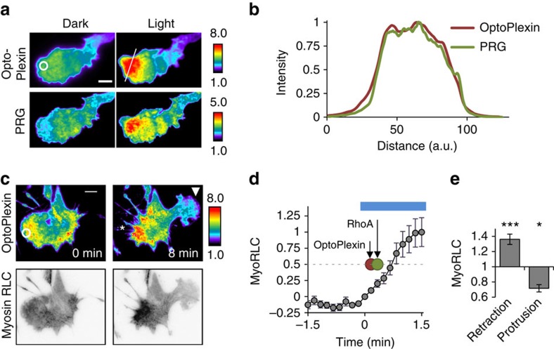
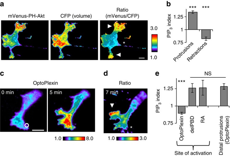
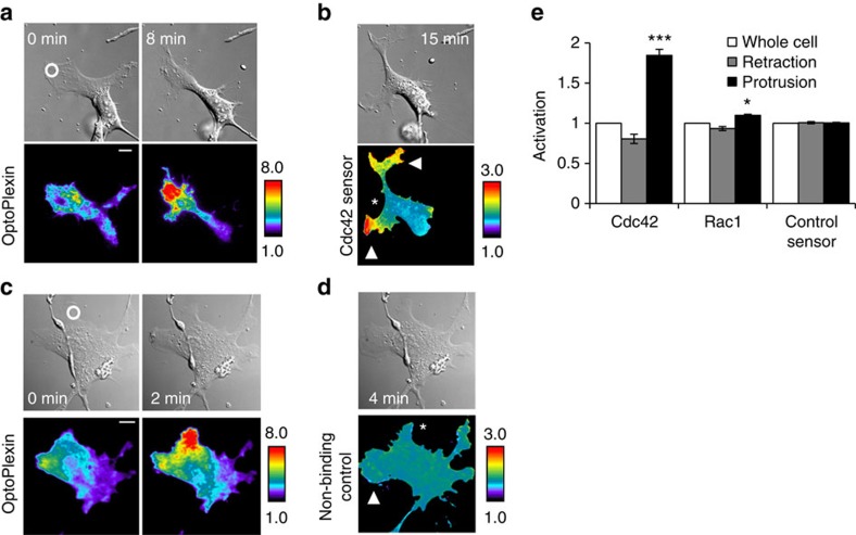
During bone remodelling, osteoclasts induce chemotaxis of osteoblasts and yet maintain spatial segregation. We show that osteoclasts express the repulsive guidance factor Semaphorin 4D and induce contact inhibition of locomotion (CIL) in osteoblasts through its receptor Plexin-B1. To examine causality and elucidate how localized Plexin-B1 stimulation may spatiotemporally coordinate its downstream targets in guiding cell migration, we develop an optogenetic tool for Plexin-B1 designated optoPlexin. Precise optoPlexin activation at the leading edge of migrating osteoblasts readily induces local retraction and, unexpectedly, distal protrusions to steer cells away. These morphological changes are accompanied by reorganization of Myosin II, PIP, adhesion and active Cdc42. We attribute the resultant repolarization to RhoA/ROCK-mediated redistribution of β-Pix, which activates Cdc42 and promotes protrusion. Thus, our data demonstrate a causal role of Plexin-B1 for CIL in osteoblasts and reveals a previously unknown effect of Semaphorin signalling on spatial distribution of an activator of cell migration.
在骨重塑过程中,破骨细胞诱导成骨细胞的趋化性,但保持空间隔离。我们表明破骨细胞表达排斥性导向因子 Semaforin 4D,并通过其受体 Plexin-B1 诱导成骨细胞的接触抑制性迁移(CIL)。为了检验因果关系并阐明局部 Plexin-B1 刺激如何在引导细胞迁移方面时空协调其下游靶标,我们开发了一种用于 Plexin-B1 的光遗传学工具,称为 optoPlexin。在迁移的成骨细胞的前缘进行精确的 optoPlexin 激活,可轻松诱导局部收缩和出乎意料的远端突起,从而引导细胞远离。这些形态变化伴随着肌球蛋白 II、PIP、黏附和活性 Cdc42 的重新排列。我们将由此产生的再极化归因于 RhoA/ROCK 介导的 β-Pix 重新分布,该分布激活 Cdc42 并促进突起。因此,我们的数据表明 Plexin-B1 在成骨细胞中的 CIL 中具有因果关系,并揭示了 Semaforin 信号对迁移激活剂空间分布的先前未知影响。