Kamimura Emi, Tanaka Shinpei, Takaba Masayuki, Tachi Keita, Baba Kazuyoshi
Department of Prosthodontics, School of Dentistry, Showa University, Tokyo, Japan.
PLoS One. 2017 Jun 21;12(6):e0179188. doi: 10.1371/journal.pone.0179188. eCollection 2017.
The aim of this study was to evaluate and compare the inter-operator reproducibility of three-dimensional (3D) images of teeth captured by a digital impression technique to a conventional impression technique in vivo.
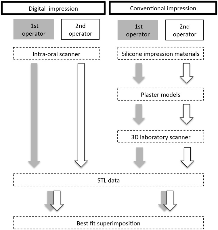
Twelve participants with complete natural dentition were included in this study. A digital impression of the mandibular molars of these participants was made by two operators with different levels of clinical experience, 3 or 16 years, using an intra-oral scanner (Lava COS, 3M ESPE). A silicone impression also was made by the same operators using the double mix impression technique (Imprint3, 3M ESPE). Stereolithography (STL) data were directly exported from the Lava COS system, while STL data of a plaster model made from silicone impression were captured by a three-dimensional (3D) laboratory scanner (D810, 3shape). The STL datasets recorded by two different operators were compared using 3D evaluation software and superimposed using the best-fit-algorithm method (least-squares method, PolyWorks, InnovMetric Software) for each impression technique. Inter-operator reproducibility as evaluated by average discrepancies of corresponding 3D data was compared between the two techniques (Wilcoxon signed-rank test).
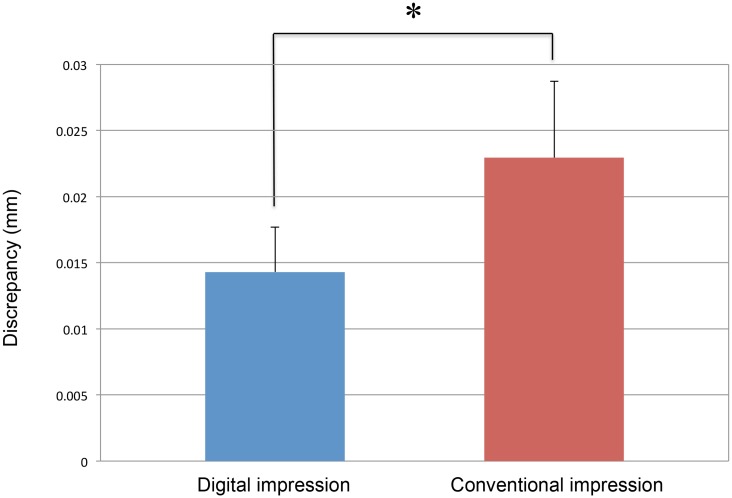
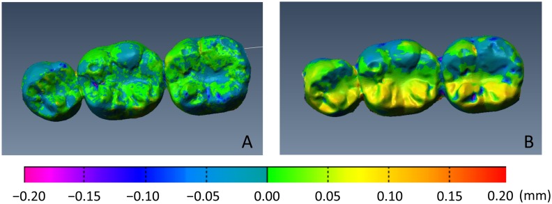
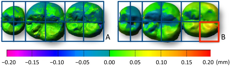
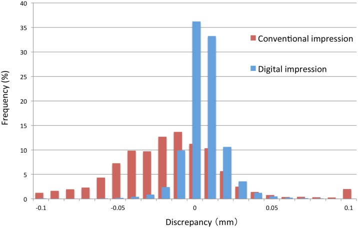
The visual inspection of superimposed datasets revealed that discrepancies between repeated digital impression were smaller than observed with silicone impression. Confirmation was forthcoming from statistical analysis revealing significantly smaller average inter-operator reproducibility using a digital impression technique (0.014± 0.02 mm) than when using a conventional impression technique (0.023 ± 0.01 mm).
The results of this in vivo study suggest that inter-operator reproducibility with a digital impression technique may be better than that of a conventional impression technique and is independent of the clinical experience of the operator.
本研究旨在评估和比较通过数字印模技术获取的牙齿三维(3D)图像与传统印模技术在体内的操作者间再现性。
本研究纳入了12名天然牙列完整的参与者。由两名临床经验水平不同(3年或16年)的操作者使用口腔内扫描仪(Lava COS,3M ESPE)对这些参与者的下颌磨牙进行数字印模。同样的操作者还使用双混印模技术(Imprint3,3M ESPE)制作了硅橡胶印模。立体光刻(STL)数据直接从Lava COS系统导出,而由硅橡胶印模制作的石膏模型的STL数据则由三维(3D)实验室扫描仪(D810,3shape)采集。使用3D评估软件比较两名不同操作者记录的STL数据集,并对每种印模技术使用最佳拟合算法方法(最小二乘法,PolyWorks,InnovMetric Software)进行叠加。通过比较两种技术对应3D数据的平均差异来评估操作者间再现性(Wilcoxon符号秩检验)。
对叠加数据集的目视检查显示,重复数字印模之间的差异小于硅橡胶印模观察到的差异。统计分析证实,使用数字印模技术时操作者间平均再现性(0.014±0.02 mm)明显小于使用传统印模技术时(0.023±0.01 mm)。
这项体内研究的结果表明,数字印模技术的操作者间再现性可能优于传统印模技术,且与操作者的临床经验无关。