Department of Organ Anatomy and Nanomedicine, Yamaguchi University Graduate School of Medicine, 1-1-1 Minami-Kogushi, Ube, Yamaguchi, 755-8505, Japan.
Division of Materials Research, Institute of Materials and Systems for Sustainability, Nagoya University, Fro-cho, Chikusa-ku, Nagoya, 464-8603, Japan.
Sci Rep. 2017 Jun 21;7(1):3953. doi: 10.1038/s41598-017-04043-7.
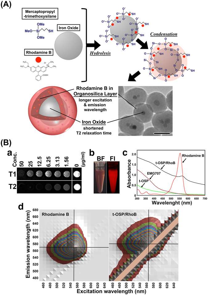
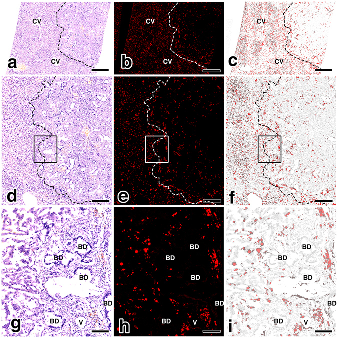
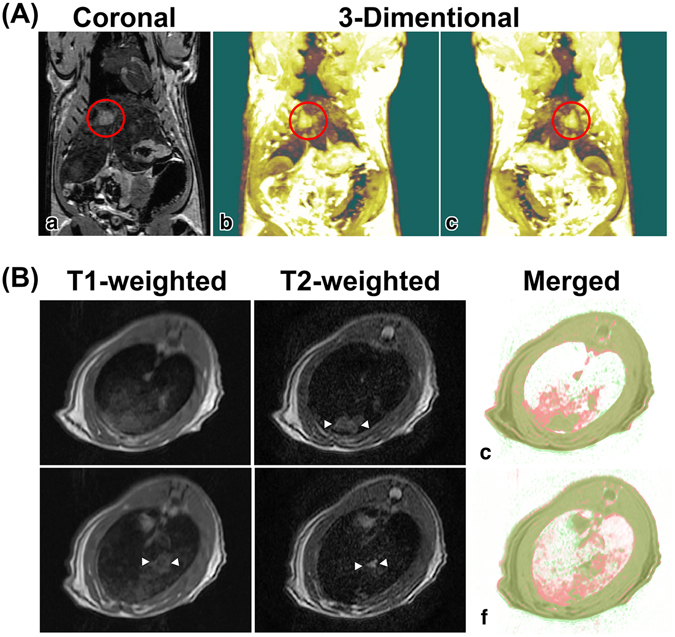
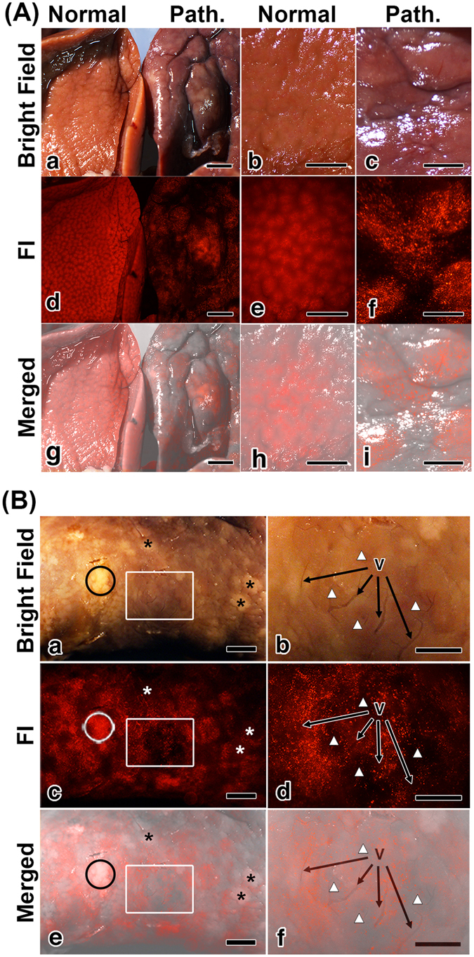
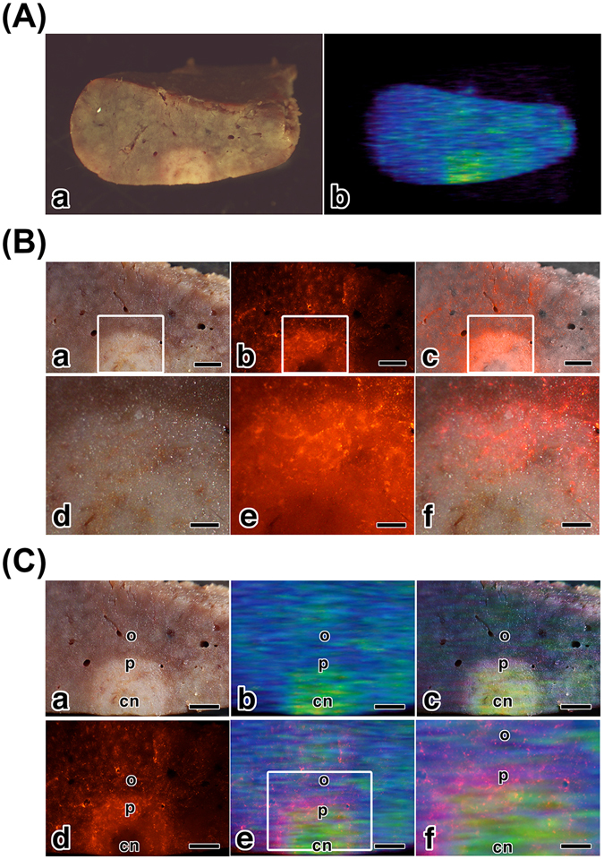
Multimodal imaging using novel multifunctional nanoparticles provides new approach to biomedical field. Thiol-organosilica nanoparticles containing iron oxide magnetic nanoparticles (MNPs) and rhodamine B (thiol OS-MNP/Rho) were applied to multimodal imaging of hepatic tumor of Long-Evans Cinnamon (LEC) rat. The magnetic resonance imaging (MRI) of LEC rats revealed tumors in the liver clearly and semi-quantitatively due to a labeling of macrophages in liver. The fluorescent imaging (FI) showed abnormal fluorescent patterns of the liver at the mesoscopic level that was between macroscopic and microscopic level. We performed correlation analysis between optical imaging including FI and MRI. We found that the labeled macrophages located specific area in the tumor tissue and influenced the tumor size on MRI. In addition histological observation showed the labeled macrophages related specific tissue in the pathological region. We demonstrated a new approach to evaluate tumor tissue at the macroscopic and microscopic level as well as mesoscopic level using multimodal imaging.
新型多功能纳米粒子的多模态成像为生物医学领域提供了新方法。含氧化铁磁性纳米粒子(MNPs)和罗丹明 B(巯基有机硅纳米粒子/Rho)的巯基有机硅纳米粒子被应用于长耳肉桂(LEC)大鼠肝肿瘤的多模态成像。磁共振成像(MRI)显示,由于肝脏中巨噬细胞的标记,大鼠肝脏中的肿瘤清晰且半定量。荧光成像(FI)显示肝在宏观和微观水平之间的介观水平上的异常荧光模式。我们对包括 FI 和 MRI 在内的光学成像进行了相关分析。我们发现,标记的巨噬细胞位于肿瘤组织的特定区域,并影响 MRI 上的肿瘤大小。此外,组织学观察显示,在病理区域中标记的巨噬细胞与特定组织有关。我们展示了一种使用多模态成像在宏观、微观和介观水平上评估肿瘤组织的新方法。