Heo Mi Hwa, Kim Hee Kyung, Lee Hansang, Kim Kyoung-Mee, Lee Jeeyun, Park Se Hoon, Park Joon Oh, Lim Ho Yeong, Kang Won Ki, Park Young Suk, Kim Seung Tae
Division of Hematology-Oncology, Department of Medicine, Samsung Medical Center, Sungkyunkwan University School of Medicine, Seoul, Korea.
Department of Pathology & Translational Genomics, Samsung Medical Center, Sungkyunkwan University School of Medicine, Seoul, Korea.
J Cancer. 2017 May 12;8(8):1395-1399. doi: 10.7150/jca.17898. eCollection 2017.
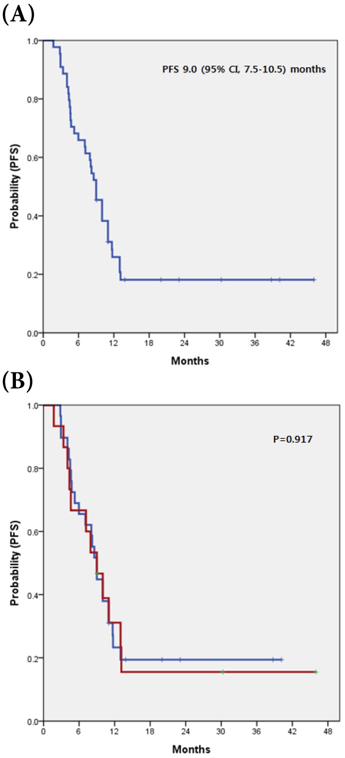
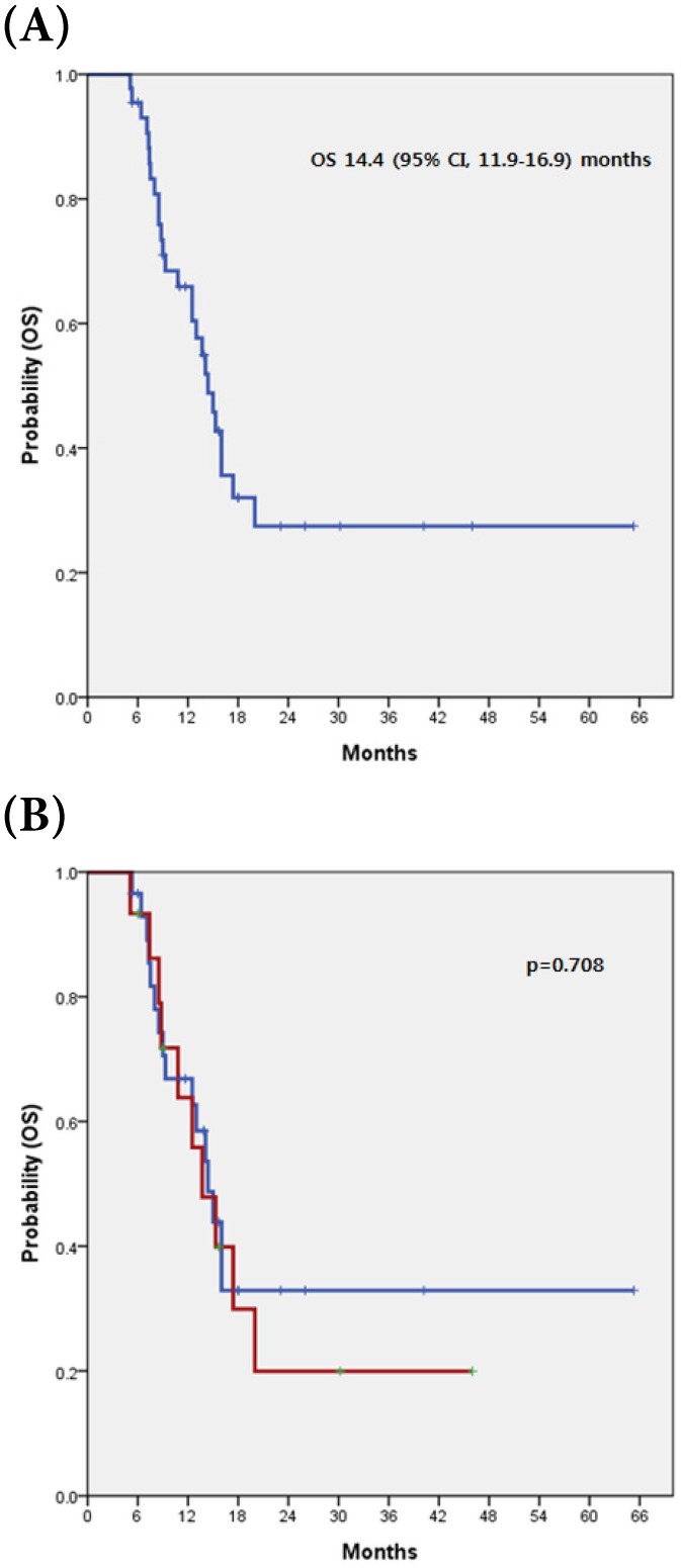
: c-MET is a proto-oncogene that encodes the tyrosine kinase receptor for hepatocyte growth factor (HGF). Activation of HGF-c-MET signaling involves cell invasiveness and evokes metastasis through direct involvement of tumor angiogenesis. However, the value of c-MET overexpression is still unknown in metastatic biliary tract cancer (BTC). : We analyzed the incidence and clinicopathologic characteristics of c-MET overexpression in advanced BTC. Moreover, we investigated the value of c-MET overexpression in predicting response to gemicitabine plus cisplatin (GC), a first line standard regimen, and as a prognostic marker in metastatic BTC. : The BTC subtype distribution (N=44) was as follows: intrahepatic cholangiocarcinoma (IHCC, n=7), extrahepatic cholangiocarcinoma (EHCC, n=25) and gallbladder cancer (GBC, n=12). Liver (52.3%) was the predominant metastatic site, followed by lymph nodes (36.4%) and bone (15.9%). Among the 44 patients analyzed for c-MET expression, 15 (34.1%) exhibited c-MET overexpression in tumor tissues. There was no significant difference in the prevalence of c-MET overexpression among primary sites in EHCC (7/25, 28.0%), IHCC (3/7, 42.9%), and GBC (5/12, 41.7%). There was also no significant correlation between specific clinicopathologic variables and c-MET expression. Comparing the tumor-response to GC according to c-MET expression (overexpression vs. non-overexpression), there was no significant difference in either RR or DCR (p=0.394 and p >0.999, respectively). The median PFS for all 44 patients was 9.00 months (95% CI, 7.5-10.5 months) and there was no significant difference for PFS between patients with c-MET overexpression and those without (p=0.917). The median OS was 14.4 months (95% CI, 11.9-16.9 months). There was no significant difference in OS between patients with c-MET overexpression compared to those without (13.7 vs. 14.4 months, respectively; p=0.708). : c-MET overexpression was detected in 34.1% of advanced BTC patients irrespective of tumor location. c-MET overexpression did not predict response to GC or survival. Further studies are needed to fully elucidate the value of c-MET overexpression as a novel biomarker in these patients.
c-MET是一种原癌基因,编码肝细胞生长因子(HGF)的酪氨酸激酶受体。HGF-c-MET信号通路的激活涉及细胞侵袭性,并通过直接参与肿瘤血管生成引发转移。然而,c-MET过表达在转移性胆管癌(BTC)中的价值仍不清楚。
我们分析了晚期BTC中c-MET过表达的发生率及临床病理特征。此外,我们研究了c-MET过表达在预测对吉西他滨联合顺铂(GC,一线标准方案)的反应以及作为转移性BTC的预后标志物方面的价值。
BTC的亚型分布(N = 44)如下:肝内胆管癌(IHCC,n = 7)、肝外胆管癌(EHCC,n = 25)和胆囊癌(GBC,n = 12)。肝脏(52.3%)是主要的转移部位,其次是淋巴结(36.4%)和骨(15.9%)。在分析c-MET表达的44例患者中,15例(34.1%)在肿瘤组织中表现出c-MET过表达。EHCC(7/25,28.0%)、IHCC(3/7,42.9%)和GBC(5/12,41.7%)原发部位中c-MET过表达的发生率无显著差异。特定临床病理变量与c-MET表达之间也无显著相关性。根据c-MET表达(过表达与未过表达)比较对GC的肿瘤反应,RR或DCR均无显著差异(分别为p = 0.394和p>0.999)。44例患者的中位PFS为9.00个月(95%CI,7.5 - 10.5个月),c-MET过表达患者与未过表达患者的PFS无显著差异(p = 0.917)。中位OS为14.4个月(95%CI,11.9 - 16.9个月)。c-MET过表达患者与未过表达患者的OS无显著差异(分别为13.7个月和14.4个月;p = 0.708)。
无论肿瘤位置如何,34.1%的晚期BTC患者检测到c-MET过表达。c-MET过表达不能预测对GC的反应或生存情况。需要进一步研究以充分阐明c-MET过表达作为这些患者新型生物标志物的价值。