Division of MR Research, Department of Radiology, Johns Hopkins University, Baltimore, Maryland, USA.
F.M. Kirby Research Center for Functional Brain Imaging, Kennedy Krieger Institute, Baltimore, Maryland, USA.
Magn Reson Med. 2017 Sep;78(3):871-880. doi: 10.1002/mrm.26799. Epub 2017 Jun 21.
To quantify amide protein transfer (APT) effects in acidic ischemic lesions and assess the spatial-temporal relationship among diffusion, perfusion, and pH deficits in acute stroke patients.
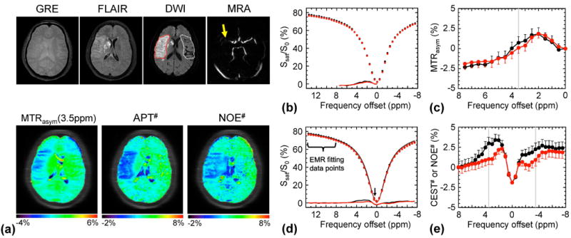
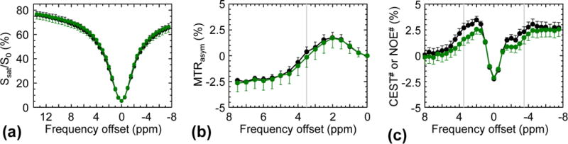
Thirty acute stroke patients were scanned at 3 T. Quantitative APT (APT ) effects in acidic ischemic lesions were measured using an extrapolated semisolid magnetization transfer reference signal technique and compared with commonly used MTR (3.5ppm) or APT-weighted parameters.
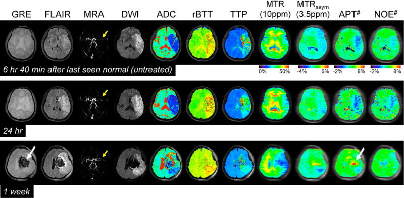
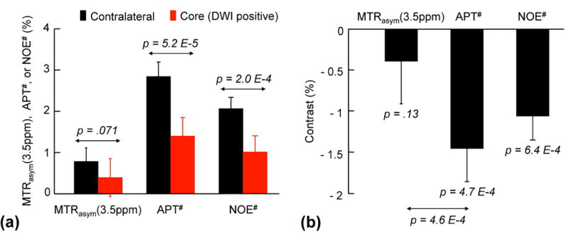
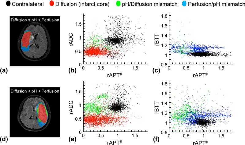
The APT images showed clear pH deficits in the ischemic lesion, whereas the MTR (3.5ppm) signals were slightly hypointense. The APT contrast between acidic ischemic lesions and normal tissue in acute stroke patients was more than three times larger than MTR (3.5ppm) contrast (-1.45 ± 0.40% for APT versus -0.39 ± 0.52% for MTR (3.5ppm), P < 4.6 × 10 ). Hypoperfused and acidic areas without an apparent diffusion coefficient abnormality were observed and assigned to an ischemic acidosis penumbra. Hypoperfused areas at normal pH were also observed and assigned to benign oligemia. Hyperintense APT signals were observed in a hemorrhage area in one case.
The quantitative APT study using the extrapolated semisolid magnetization transfer reference signal approach enhances APT MRI sensitivity to pH compared with conventional APT-weighted MRI, allowing more reliable delineation of an ischemic acidosis in the penumbra. Magn Reson Med 78:871-880, 2017. © 2017 International Society for Magnetic Resonance in Medicine.
量化酸性缺血病灶中的酰胺蛋白转移(APT)效应,并评估急性脑卒中患者扩散、灌注和 pH 缺陷之间的时空关系。
对 30 例急性脑卒中患者在 3T 下进行扫描。使用外推半固态磁化传递参考信号技术测量酸性缺血性病变中的定量 APT(APT)效应,并与常用的 MTR(3.5ppm)或 APT 加权参数进行比较。
APT 图像显示缺血性病变中存在明显的 pH 缺陷,而 MTR(3.5ppm)信号则略有低信号。与 MTR(3.5ppm)相比,急性脑卒中患者酸性缺血性病变与正常组织之间的 APT 对比度大 3 倍以上(APT:-1.45±0.40%,MTR(3.5ppm):-0.39±0.52%,P<4.6×10⁻⁴)。观察到存在灌注不足和无明显扩散系数异常的酸性区域,并将其分配到缺血性酸中毒半影区。也观察到 pH 正常的灌注不足区域,并将其分配到良性低氧血症。在一个病例中,观察到出血区域有高亮 APT 信号。
与传统的 APT 加权 MRI 相比,使用外推半固态磁化传递参考信号方法的定量 APT 研究增强了 APT MRI 对 pH 的灵敏度,从而更可靠地描绘了半影区的缺血性酸中毒。磁共振医学杂志 78:871-880, 2017。© 2017 国际磁共振学会。