Roberts Thomas A, Price Anthony N, Jackson Laurence H, Taylor Valerie, David Anna L, Lythgoe Mark F, Stuckey Daniel J
Centre for Advanced Biomedical Imaging, University College London, London, UK.
Division of Imaging Sciences and Biomedical Engineering, King's College London, London, UK.
NMR Biomed. 2017 Oct;30(10). doi: 10.1002/nbm.3763. Epub 2017 Jun 23.
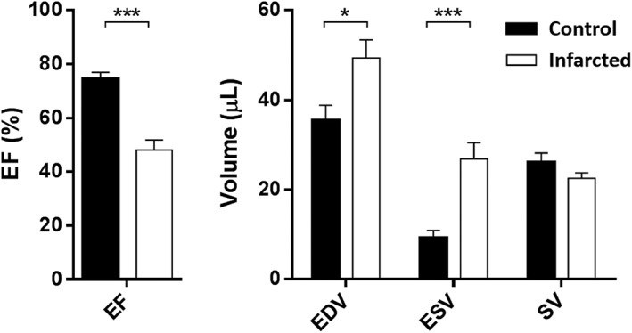
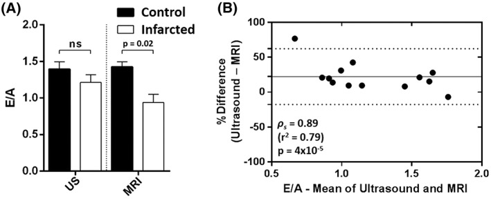
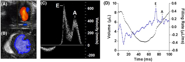
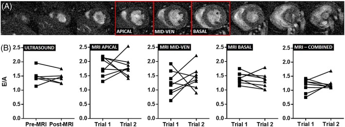
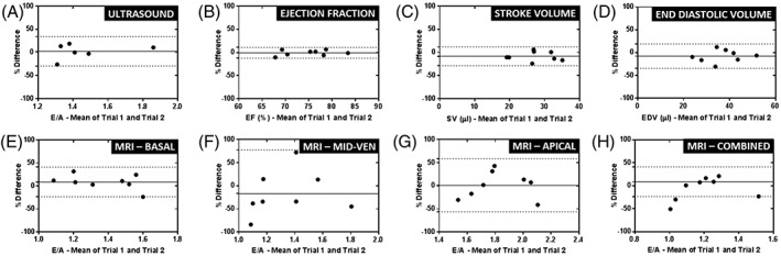
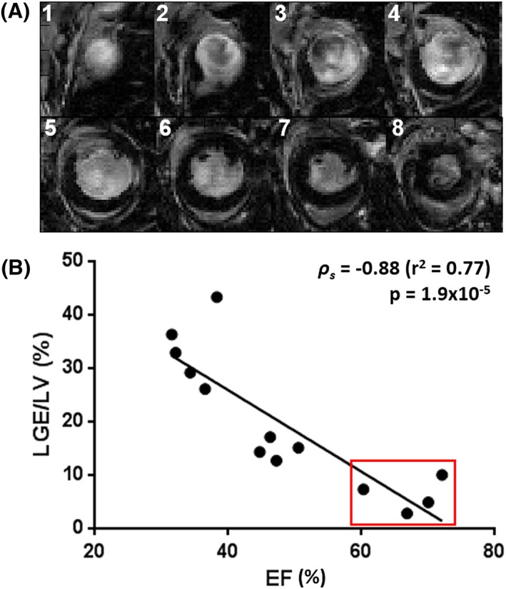
Diastolic dysfunction is a sensitive early indicator of heart failure and can provide additional data to conventional measures of systolic function. Transmitral Doppler ultrasound, which measures the one-dimensional flow of blood through the mitral valve, is currently the preferred method for the measurement of diastolic function, but the measurement of the left ventricular volume changes using high-temporal-resolution cinematic magnetic resonance imaging (CINE MRI) is an alternative approach which is emerging as a potentially more robust and user-independent technique. Here, we investigated the performance of high-temporal-resolution CINE MRI and compared it with ultrasound for the detection of diastolic dysfunction in a mouse model of myocardial infarction. An in-house, high-temporal-resolution, retrospectively gated CINE sequence was developed with a temporal resolution of 1 ms. Diastolic function in mice was assessed using a custom-made, open-source reconstruction package. Early (E) and late (A) left ventricular filling phases were easily identifiable, and these measurements were compared directly with high-frequency, pulsed-wave, Doppler ultrasound measurements of mitral valve inflow. A repeatability study established that high-temporal-resolution CINE MRI and Doppler ultrasound showed comparable accuracy when measuring E/A in normal control mice. However, when applied in a mouse model of myocardial infarction, high-temporal-resolution CINE MRI indicated diastolic heart failure (E/A = 0.94 ± 0.11), whereas ultrasound falsely detected normal cardiac function (E/A = 1.21 ± 0.11). The addition of high-temporal-resolution CINE MRI to preclinical imaging studies enhances the library of sequences available to cardiac researchers and potentially identifies diastolic heart failure early in disease progression.
舒张功能障碍是心力衰竭的一个敏感早期指标,可为收缩功能的传统测量方法提供额外数据。经二尖瓣多普勒超声可测量通过二尖瓣的一维血流,目前是测量舒张功能的首选方法,但使用高时间分辨率电影磁共振成像(CINE MRI)测量左心室容积变化是一种替代方法,正逐渐成为一种可能更可靠且不依赖用户的技术。在此,我们研究了高时间分辨率CINE MRI的性能,并将其与超声在心肌梗死小鼠模型中检测舒张功能障碍的情况进行了比较。我们开发了一种内部的、高时间分辨率、回顾性门控的CINE序列,时间分辨率为1毫秒。使用定制的开源重建软件包评估小鼠的舒张功能。左心室早期(E)和晚期(A)充盈期很容易识别,并将这些测量结果直接与二尖瓣流入的高频脉冲波多普勒超声测量结果进行比较。一项重复性研究表明,在测量正常对照小鼠的E/A时,高时间分辨率CINE MRI和多普勒超声显示出相当的准确性。然而,当应用于心肌梗死小鼠模型时,高时间分辨率CINE MRI显示舒张性心力衰竭(E/A = 0.94 ± 0.11),而超声错误地检测到心脏功能正常(E/A = 1.21 ± 0.11)。将高时间分辨率CINE MRI添加到临床前成像研究中,可增加心脏研究人员可用的序列库,并有可能在疾病进展早期识别出舒张性心力衰竭。