Kang Chengrong, Wei Limin, Song Bin, Chen Liangjiao, Liu Jia, Deng Bin, Pan Xuan, Shao Longquan
Department of Stomatology, Nanfang Hospital, Southern Medical University.
Department of Stomatology, The First Affiliated Hospital of Guangdong Pharmaceutical University.
Int J Nanomedicine. 2017 Jun 7;12:4323-4333. doi: 10.2147/IJN.S136281. eCollection 2017.
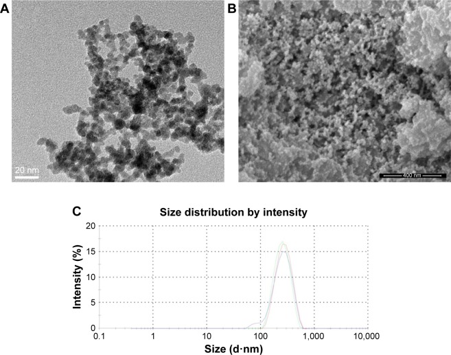
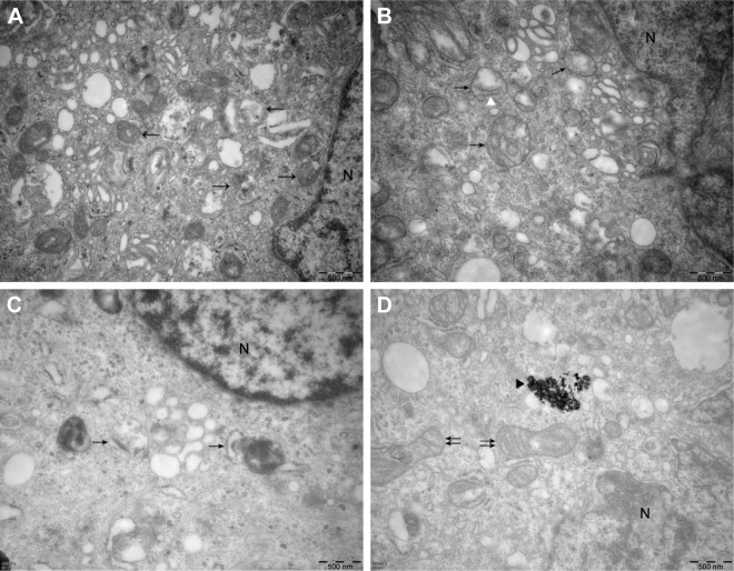
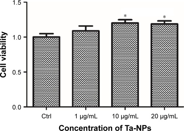
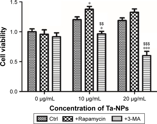
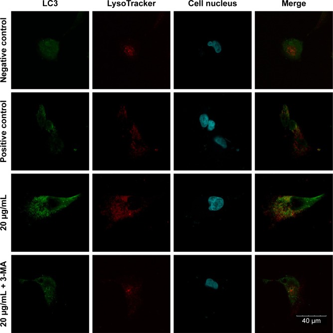
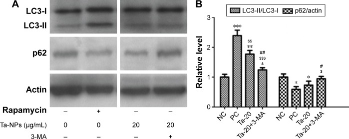
Porous tantalum (Ta) implants are highly corrosion resistant and biocompatible, and they possess significantly better initial stability than that of conventional titanium (Ti) implants. During loading wear, Ta nanoparticles (Ta-NPs) that were deposited on the surface of a porous Ta implant are inevitably released and come into direct contact with peri-implant osteoblasts. The wear debris may influence cell behavior and implant stabilization. However, the interaction of Ta-NPs with osteoblasts has not been clearly investigated. This study aimed to investigate the effect of Ta-NPs on cell proliferation and their underlying mechanism. The Cell Counting Kit-8 (CCK-8) assay was used to measure the cell viability of MC3T3-E1 mouse osteoblasts and showed that Ta-NP treatment could increase cell viability. Then, confocal microscopy, Western blotting, and transmission electron microscopy were used to confirm the autophagy induced by Ta-NPs, and evidence of autophagy induction was observed as positive LC3 puncta, high-LC3-II expression, and autophagic vesicle ultrastructures. The CCK-8 assay revealed that the cell viability was further increased and decreased by the application of an autophagy inducer and inhibitor, respectively. In addition, pre-treatment with autophagy inhibitor 3-methyladenine (3-MA) inhibited the Ta-NP-induced autophagy. These results indicate that the Ta-NPs can promote cell proliferation, that an autophagy inducer can further strengthen this effect and that an autophagy inhibitor can weaken this effect. In conclusion, autophagy was involved in Ta-NP-induced cell proliferation and had a promoting effect.
多孔钽(Ta)植入物具有高度的耐腐蚀性和生物相容性,并且它们具有比传统钛(Ti)植入物明显更好的初始稳定性。在加载磨损过程中,沉积在多孔Ta植入物表面的Ta纳米颗粒(Ta-NPs)不可避免地释放出来,并与植入物周围的成骨细胞直接接触。磨损碎片可能会影响细胞行为和植入物的稳定性。然而,Ta-NPs与成骨细胞之间的相互作用尚未得到明确研究。本研究旨在探讨Ta-NPs对细胞增殖的影响及其潜在机制。使用细胞计数试剂盒-8(CCK-8)测定法来测量MC3T3-E1小鼠成骨细胞的细胞活力,结果表明Ta-NP处理可提高细胞活力。然后,使用共聚焦显微镜、蛋白质印迹法和透射电子显微镜来确认Ta-NPs诱导的自噬,观察到自噬诱导的证据为阳性LC3斑点、高LC3-II表达和自噬泡超微结构。CCK-8测定法显示,分别应用自噬诱导剂和抑制剂后,细胞活力进一步增加和降低。此外,用自噬抑制剂3-甲基腺嘌呤(3-MA)预处理可抑制Ta-NP诱导的自噬。这些结果表明,Ta-NPs可以促进细胞增殖,自噬诱导剂可以进一步增强这种作用,而自噬抑制剂可以减弱这种作用。总之,自噬参与了Ta-NP诱导的细胞增殖并具有促进作用。