Jayakumar Asha, Bothwell Alfred L M
Department of Immunobiology, Yale University School of Medicine, New Haven, CT 06520.
Department of Immunobiology, Yale University School of Medicine, New Haven, CT 06520.
Neoplasia. 2017 Aug;19(8):595-605. doi: 10.1016/j.neo.2017.04.006. Epub 2017 Jun 24.
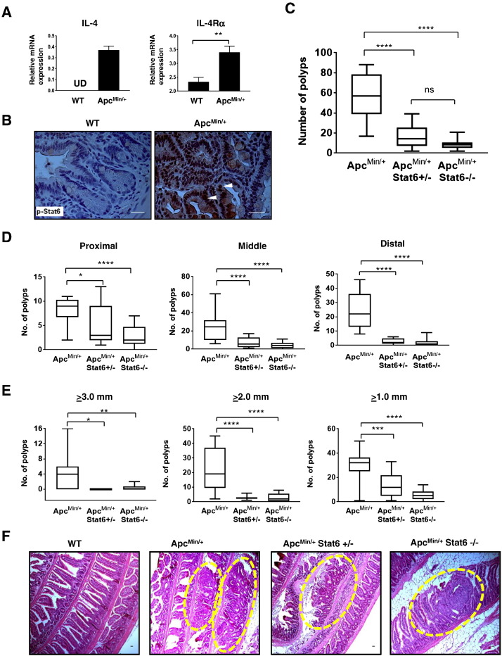
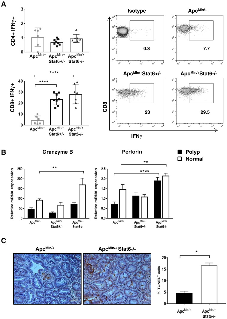
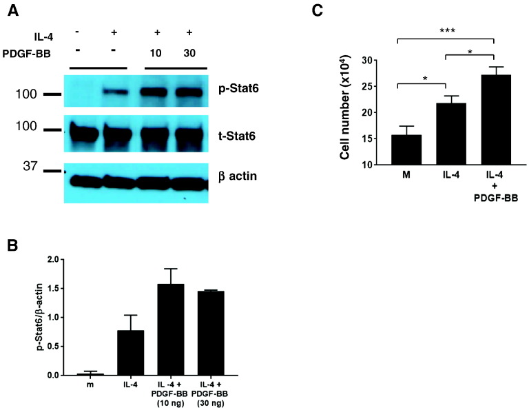
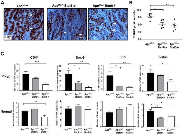
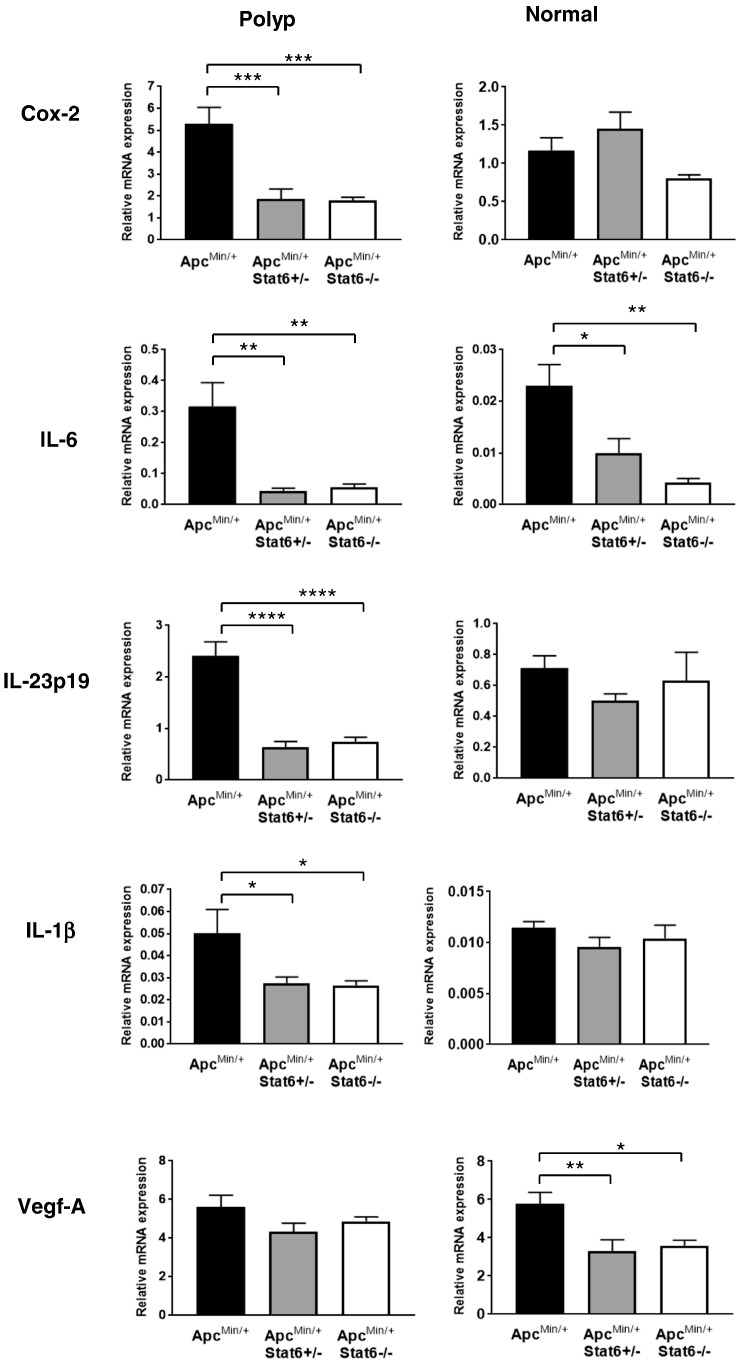
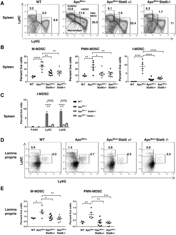
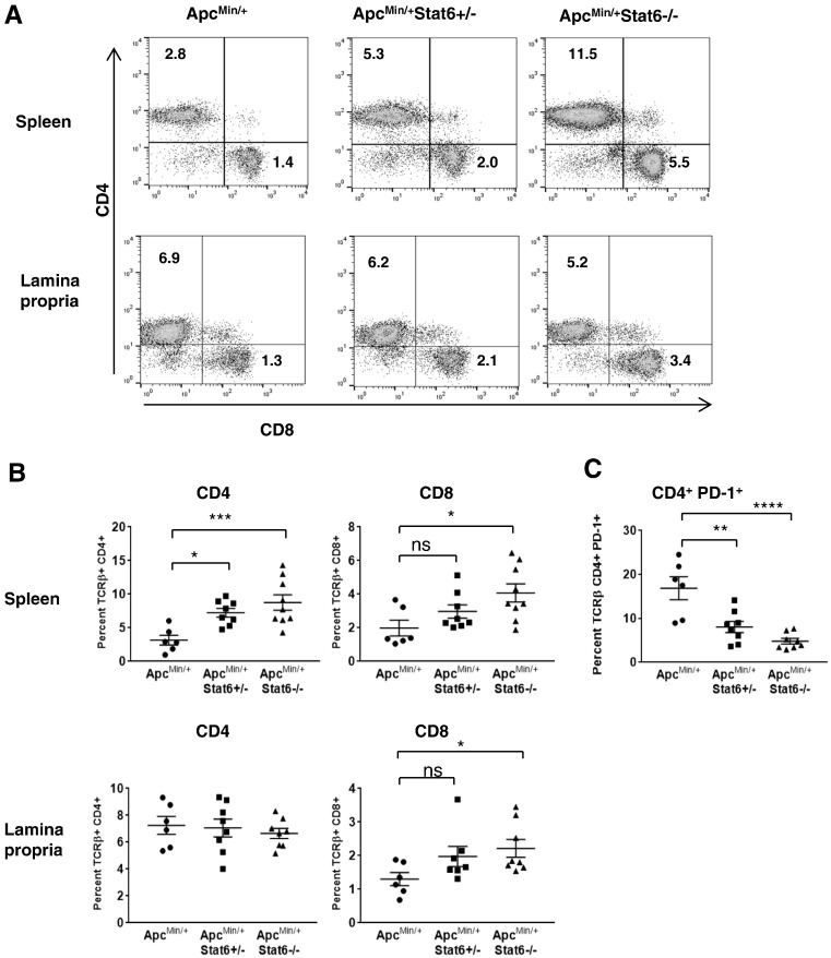
Intestinal tumorigenesis in the ApcMin/+ model is initiated by aberrant activation of Wnt pathway. Increased IL-4 expression in human colorectal cancer tissue and growth of colon cancer cell lines implied that IL-4-induced Stat6-mediated tumorigenic signaling likely contributes to intestinal tumor progression in ApcMin/+ mice. Stat6 also appears to promote expansion of myeloid-derived suppressor cells (MDSCs) cells. MDSCs promote polyp formation in the ApcMin/+ model. Hence, Stat6 could have a broad role in coordinating both polyp cell proliferation and MDSC expansion. We found that IL-4-induced Stat6-mediated proliferation of intestinal epithelial cells is augmented by platelet-derived growth factor-BB, a tumor-promoting growth factor. To determine whether polyp progression in ApcMin/+ mice is dependent on Stat6 signaling, we disrupted Stat6 in this model. Total polyps in the small intestine were fewer in ApcMin/+ mice lacking Stat6. Furthermore, proliferation of polyp epithelial cells was reduced, indicating that Stat6 in part controlled polyp formation. Stat6 also promoted expansion of MDSCs in the spleen and lamina propria of ApcMin/+ mice, implying regulation of antitumor T-cell response. More CD8 cells and reduced PD-1 expression on CD4 cells correlated with reduced polyps. In addition, a strong CD8-mediated cytotoxic response led to killing of tumor cells in Stat6-deficient ApcMin/+ mice. Therefore, these findings show that Stat6 has an oncogenic role in intestinal tumorigenesis by promoting polyp cell proliferation and immunosuppressive mediators, and preventing an active cytotoxic process.
ApcMin/+模型中的肠道肿瘤发生是由Wnt信号通路的异常激活引发的。人类结直肠癌组织中白细胞介素-4(IL-4)表达增加以及结肠癌细胞系的生长表明,IL-4诱导的Stat6介导的致瘤信号可能促成了ApcMin/+小鼠的肠道肿瘤进展。Stat6似乎还能促进髓源性抑制细胞(MDSCs)的扩增。MDSCs在ApcMin/+模型中促进息肉形成。因此,Stat6在协调息肉细胞增殖和MDSC扩增方面可能具有广泛作用。我们发现,血小板衍生生长因子-BB(一种促肿瘤生长因子)增强了IL-4诱导的Stat6介导的肠道上皮细胞增殖。为了确定ApcMin/+小鼠的息肉进展是否依赖于Stat6信号,我们在该模型中破坏了Stat6。缺乏Stat6的ApcMin/+小鼠小肠中的息肉总数较少。此外,息肉上皮细胞的增殖减少,表明Stat6部分控制息肉形成。Stat6还促进了ApcMin/+小鼠脾脏和固有层中MDSCs的扩增,这意味着对抗肿瘤T细胞反应的调节。更多的CD8细胞以及CD4细胞上PD-1表达的降低与息肉减少相关。此外,强烈的CD8介导的细胞毒性反应导致Stat6缺陷的ApcMin/+小鼠中的肿瘤细胞被杀伤。因此,这些发现表明,Stat6通过促进息肉细胞增殖和免疫抑制介质以及阻止活跃的细胞毒性过程,在肠道肿瘤发生中具有致癌作用。