Department of Ophthalmology, Eye Institute of China PLA, Xijing Hospital, Fourth Military Medical University, Xi'an, China.
Department of Ophthalmology, Jinling Hospital, Nanjing, China.
J Cell Mol Med. 2017 Dec;21(12):3467-3480. doi: 10.1111/jcmm.13256. Epub 2017 Jun 29.
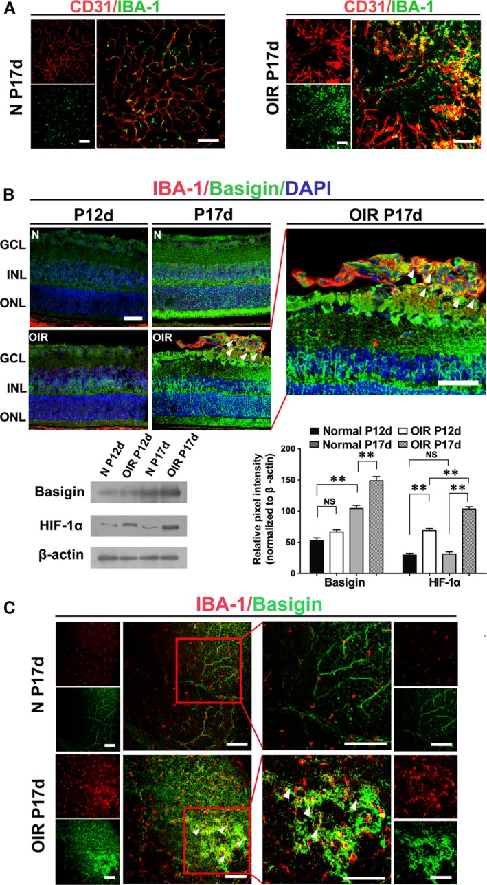
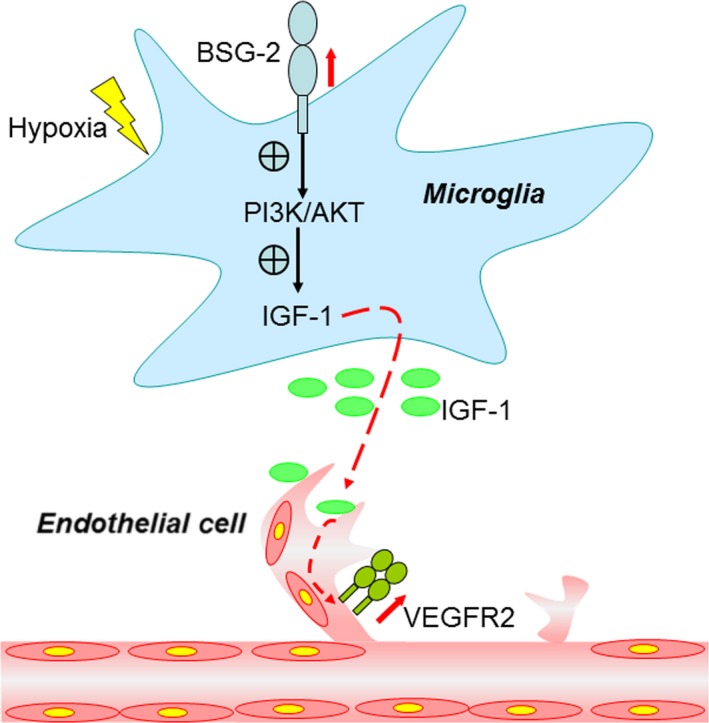
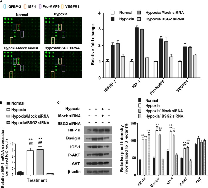
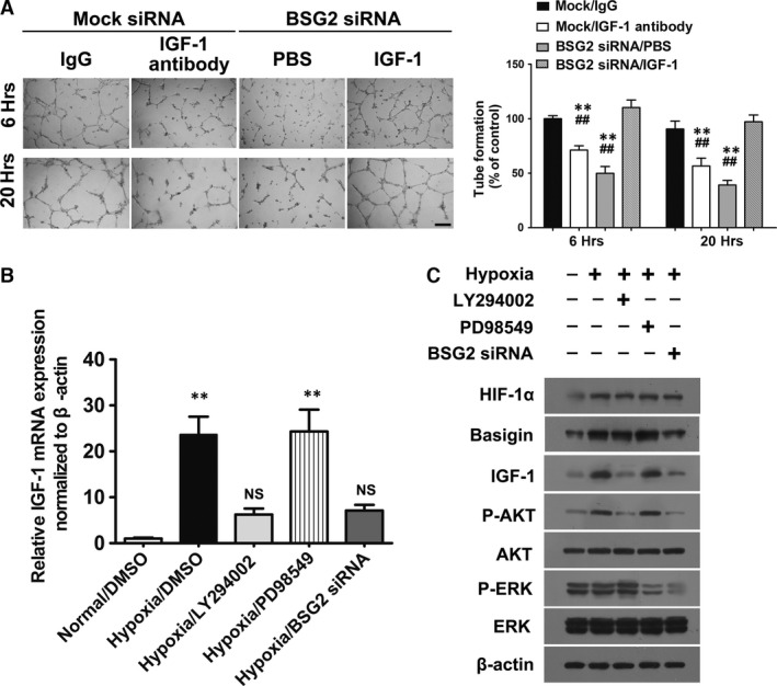
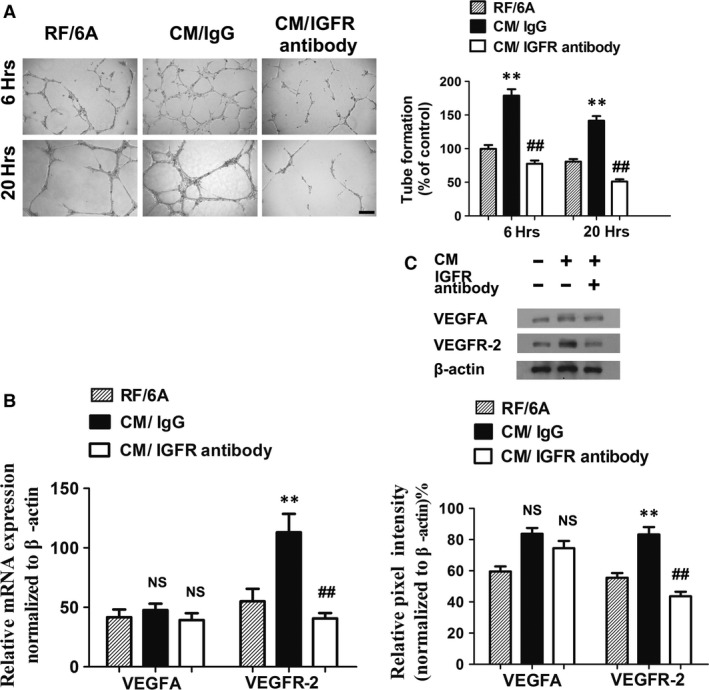
Retinal microglia cells contribute to vascular angiogenesis and vasculopathy induced by relative hypoxia. However, its concrete molecular mechanisms in shaping retinal angiogenesis have not been elucidated. Basigin, being involved in tumour neovasculogenesis, is explored to exert positive effects on retinal angiogenesis induced by microglia. Therefore, we set out to investigate the expression of basigin using a well-characterized mouse model of oxygen-induced retinopathy, which recapitulated hypoxia-induced aberrant neovessel growth. Our results elucidate that basigin is overexpressed in microglia, which accumulating in retinal angiogenic sprouts. In vitro, conditioned media from microglia BV2 under hypoxia treatment increase migration and tube formation of retinal capillary endothelia cells, compared with media from normoxic condition. The angiogenic capacity of BV2 is inhibited after basigin knockdown by small interfering RNAs. A new molecular mechanism for high angiogenic capacity, whereby microglia cells release basigin via up-regulation of PI3K-AKT and IGF-1 pathway to induce angiogenesis is unveiled. Collectively, our results demonstrate that basigin from hypoxic microglia plays a pivotal pro-angiogenic role, providing new insights into microglia-promoting retinal angiogenesis.
视网膜小胶质细胞有助于相对缺氧引起的血管生成和血管病变。然而,其在塑造视网膜血管生成方面的具体分子机制尚未阐明。Basigin 参与肿瘤新生血管生成,被探索对小胶质细胞诱导的视网膜血管生成发挥积极作用。因此,我们使用一种经过充分表征的氧诱导视网膜病变小鼠模型来研究 basigin 的表达,该模型重现了缺氧诱导的异常新生血管生长。我们的结果阐明了 basigin 在小胶质细胞中过度表达,这些小胶质细胞聚集在视网膜血管生成芽中。在体外,与正常氧条件下的培养基相比,缺氧处理的小胶质细胞 BV2 的条件培养基增加了视网膜毛细血管内皮细胞的迁移和管形成。用小干扰 RNA 敲低 basigin 后,BV2 的血管生成能力受到抑制。揭示了一种新的高血管生成能力的分子机制,即小胶质细胞通过上调 PI3K-AKT 和 IGF-1 通路释放 basigin 来诱导血管生成。总之,我们的结果表明,缺氧小胶质细胞中的 basigin 发挥着关键的促血管生成作用,为小胶质细胞促进视网膜血管生成提供了新的见解。