Krasnow Stephanie M, Knoll J Gabriel, Verghese Santhosh Chakkaramakkil, Levasseur Peter R, Marks Daniel L
Department of Pediatrics, Papé Family Pediatric Research Institute, Oregon Health & Science University, Portland, Oregon, 97239, USA.
Oregon Health & Science University, Mail Code L481, 3181 SW Sam Jackson Park Rd., Portland, OR, 97239, USA.
J Neuroinflammation. 2017 Jul 1;14(1):133. doi: 10.1186/s12974-017-0908-4.
During acute infections and chronic illnesses, the pro-inflammatory cytokine interleukin-1β (IL-1β) acts within the brain to elicit metabolic derangements and sickness behaviors. It is unknown which cells in the brain are the proximal targets for IL-1β with respect to the generation of these illness responses. We performed a series of in vitro experiments to (1) investigate which brain cell populations exhibit inflammatory responses to IL-1β and (2) examine the interactions between different IL-1β-responsive cell types in various co-culture combinations.
We treated primary cultures of murine brain microvessel endothelial cells (BMEC), astrocytes, and microglia with PBS or IL-1β, and then performed qPCR to measure inflammatory gene expression or immunocytochemistry to evaluate nuclear factor kappa-light-chain-enhancer of activated B cells (NF-κB) activation. To evaluate whether astrocytes and/or BMEC propagate inflammatory signals to microglia, we exposed microglia to astrocyte-conditioned media and co-cultured endothelial cells and glia in transwells. Treatment groups were compared by Student's t tests or by ANOVA followed by Bonferroni-corrected t tests.
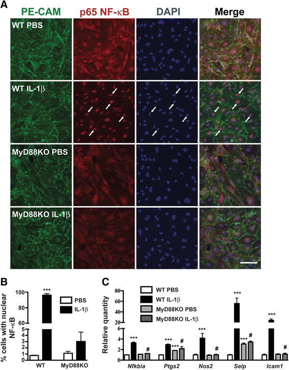
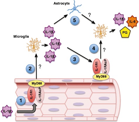
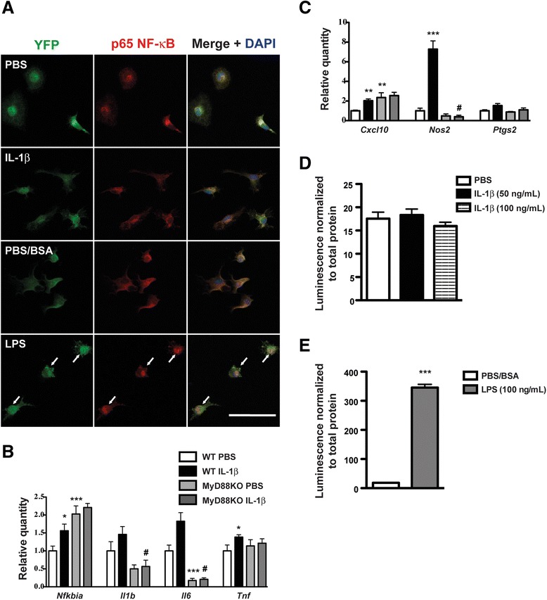
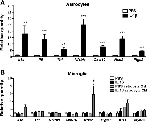
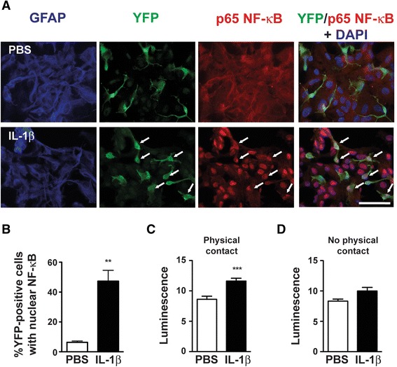
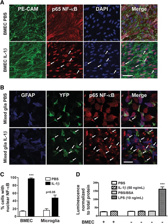
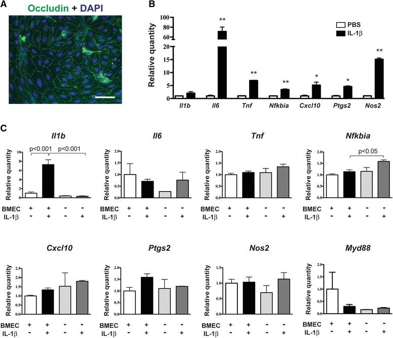
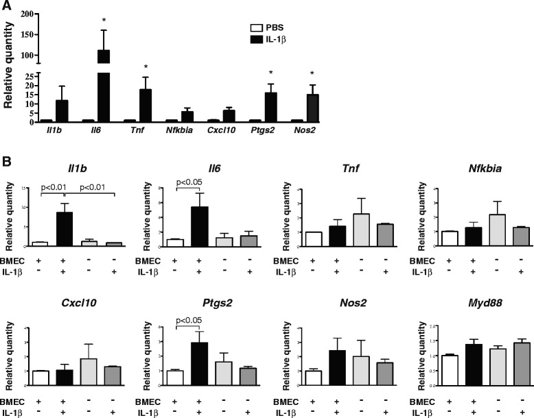
IL-1β increased inflammatory gene expression and NF-κB activation in primary murine-mixed glia, enriched astrocyte, and BMEC cultures. Although IL-1β elicited minimal changes in inflammatory gene expression and did not induce the nuclear translocation of NF-κB in isolated microglia, these cells were more robustly activated by IL-1β when co-cultured with astrocytes and/or BMEC. We observed a polarized endothelial response to IL-1β, because the application of IL-1β to the abluminal endothelial surface produced a more complex microglial inflammatory response than that which occurred following luminal IL-1β exposure.
Inflammatory signals are detected, amplified, and propagated through the CNS via a sequential and reverberating signaling cascade involving communication between brain endothelial cells and glia. We propose that the brain's innate immune response differs depending upon which side of the blood-brain barrier the inflammatory stimulus arises, thus allowing the brain to respond differently to central vs. peripheral inflammatory insults.
在急性感染和慢性疾病期间,促炎细胞因子白细胞介素-1β(IL-1β)在脑内发挥作用,引发代谢紊乱和疾病行为。关于这些疾病反应的产生,脑内哪些细胞是IL-1β的近端靶点尚不清楚。我们进行了一系列体外实验,以(1)研究哪些脑细胞群体对IL-1β表现出炎症反应,以及(2)检查不同IL-1β反应性细胞类型在各种共培养组合中的相互作用。
我们用磷酸盐缓冲液(PBS)或IL-1β处理小鼠脑微血管内皮细胞(BMEC)、星形胶质细胞和小胶质细胞的原代培养物,然后进行定量聚合酶链反应(qPCR)以测量炎症基因表达,或进行免疫细胞化学以评估活化B细胞的核因子κB(NF-κB)激活。为了评估星形胶质细胞和/或BMEC是否将炎症信号传递给小胶质细胞,我们将小胶质细胞暴露于星形胶质细胞条件培养基中,并在Transwell小室中共培养内皮细胞和神经胶质细胞。通过学生t检验或方差分析(ANOVA),然后进行Bonferroni校正的t检验来比较治疗组。
IL-1β增加了原代小鼠混合神经胶质细胞、富集的星形胶质细胞和BMEC培养物中的炎症基因表达和NF-κB激活。虽然IL-1β在分离的小胶质细胞中引起炎症基因表达的最小变化,并且没有诱导NF-κB的核转位,但当与星形胶质细胞和/或BMEC共培养时,这些细胞被IL-1β更强烈地激活。我们观察到内皮细胞对IL-1β的极化反应,因为将IL-1β应用于无腔内皮表面比应用于腔面IL-1β暴露后产生更复杂的小胶质细胞炎症反应。
炎症信号通过涉及脑内皮细胞和神经胶质细胞之间通信的顺序和回响信号级联在中枢神经系统中被检测、放大和传播。我们提出,脑的固有免疫反应因炎症刺激出现在血脑屏障的哪一侧而有所不同,从而使脑对中枢性与外周性炎症损伤做出不同反应。