Wellcome Centre for Integrative Neuroscience (WIN) - Oxford Centre for Functional Magnetic Resonance Imaging of the Brain (FMRIB), University of Oxford, UK.
Wellcome Centre for Integrative Neuroscience (WIN) - Oxford Centre for Functional Magnetic Resonance Imaging of the Brain (FMRIB), University of Oxford, UK.
Neuroimage. 2017 Sep;158:205-218. doi: 10.1016/j.neuroimage.2017.06.050. Epub 2017 Jun 29.
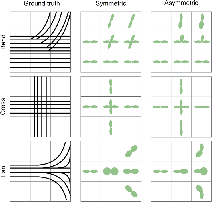
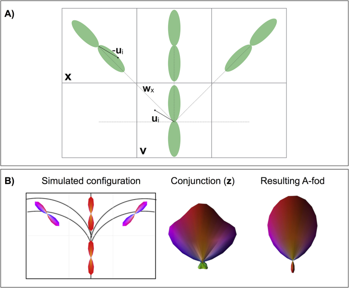
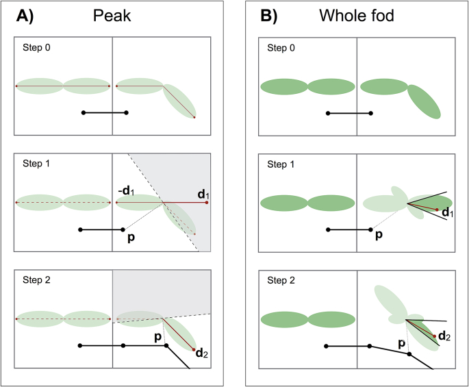
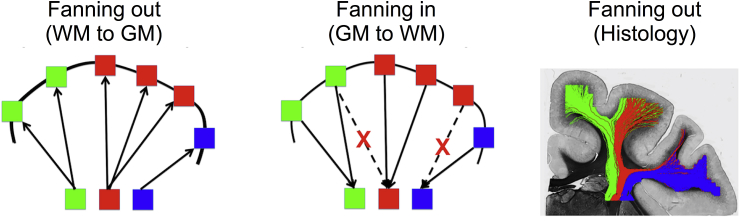
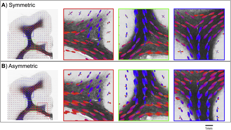
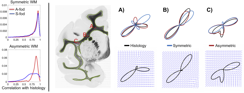
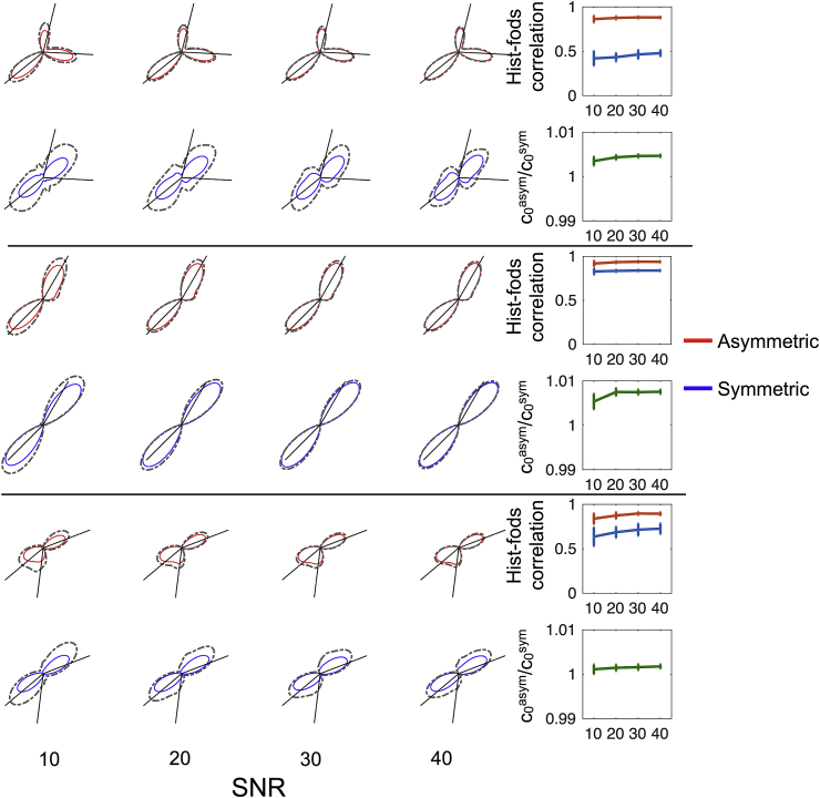
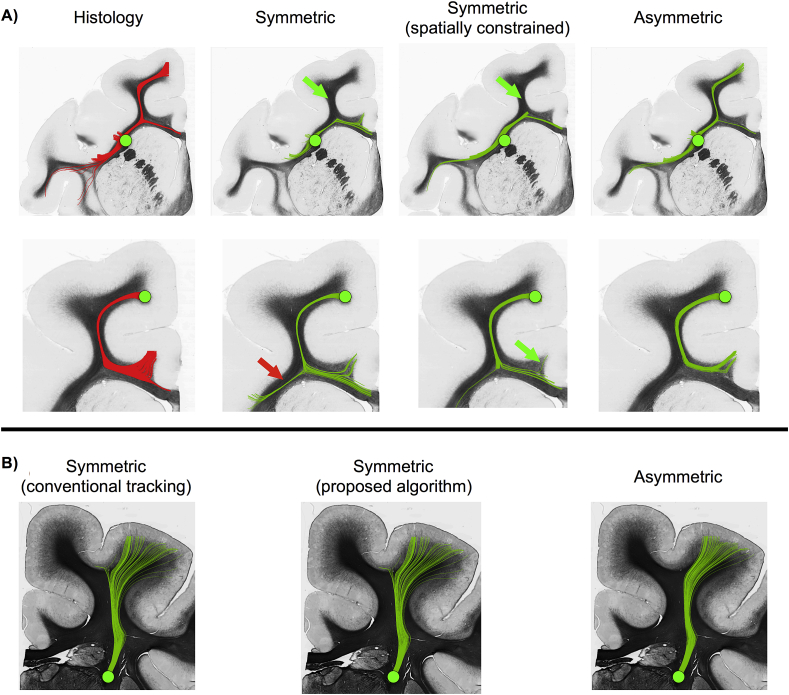
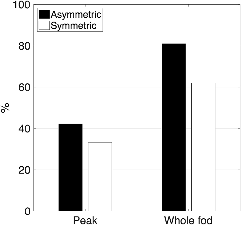
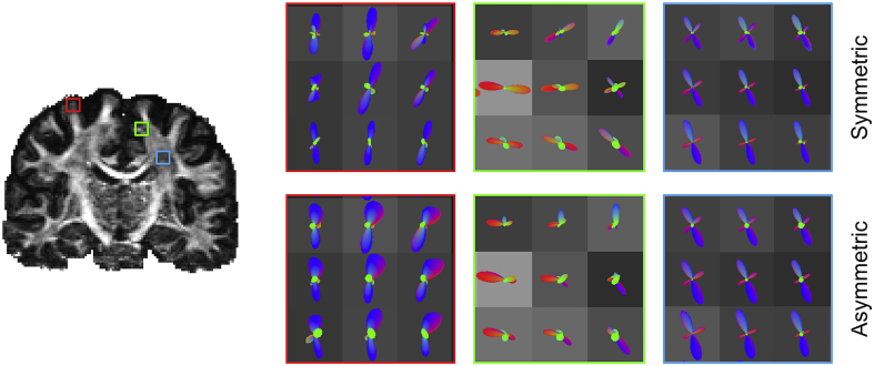
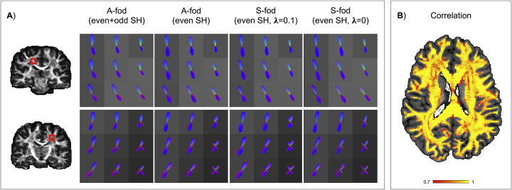
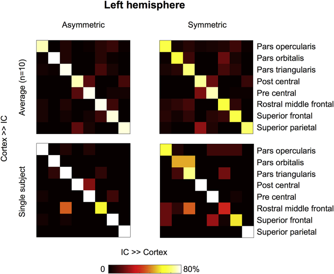
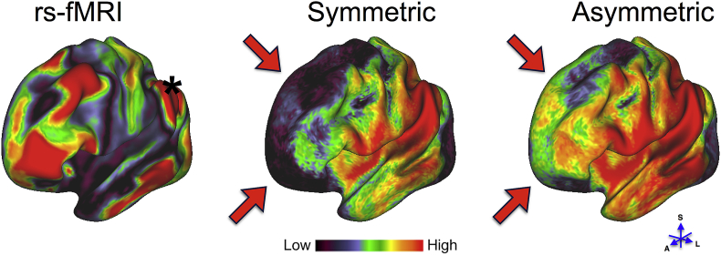
Diffusion MRI allows us to make inferences on the structural organisation of the brain by mapping water diffusion to white matter microstructure. However, such a mapping is generally ill-defined; for instance, diffusion measurements are antipodally symmetric (diffusion along x and -x are equal), whereas the distribution of fibre orientations within a voxel is generally not symmetric. Therefore, different sub-voxel patterns such as crossing, fanning, or sharp bending, cannot be distinguished by fitting a voxel-wise model to the signal. However, asymmetric fibre patterns can potentially be distinguished once spatial information from neighbouring voxels is taken into account. We propose a neighbourhood-constrained spherical deconvolution approach that is capable of inferring asymmetric fibre orientation distributions (A-fods). Importantly, we further design and implement a tractography algorithm that utilises the estimated A-fods, since the commonly used streamline tractography paradigm cannot directly take advantage of the new information. We assess performance using ultra-high resolution histology data where we can compare true orientation distributions against sub-voxel fibre patterns estimated from down-sampled data. Finally, we explore the benefits of A-fods-based tractography using in vivo data by evaluating agreement of tractography predictions with connectivity estimates made using different in-vivo modalities. The proposed approach can reliably estimate complex fibre patterns such as sharp bending and fanning, which voxel-wise approaches cannot estimate. Moreover, histology-based and in-vivo results show that the new framework allows more accurate tractography and reconstruction of maps quantifying (symmetric and asymmetric) fibre complexity.
扩散磁共振成像(Diffusion MRI)通过将水分子扩散映射到白质微观结构,使我们能够对大脑的结构组织进行推断。然而,这种映射通常是不确定的;例如,扩散测量是反向对称的(沿着 x 和-x 的扩散相等),而体素内纤维方向的分布通常是不对称的。因此,不同的亚体素模式,如交叉、扇形或急剧弯曲,不能通过将体素模型拟合到信号来区分。然而,一旦考虑到相邻体素的空间信息,就有可能区分不对称的纤维模式。我们提出了一种邻域约束的球分解方法,能够推断出不对称的纤维方向分布(A-fods)。重要的是,我们进一步设计并实现了一种跟踪算法,利用估计的 A-fods,因为常用的流线跟踪范式不能直接利用新信息。我们使用超高分辨率组织学数据来评估性能,在这些数据中,我们可以将真实的方向分布与从下采样数据中估计的亚体素纤维模式进行比较。最后,我们通过评估基于 A-fods 的跟踪与使用不同体内模态进行的连接估计的跟踪预测的一致性,探索了基于体内数据的 A-fods 跟踪的好处。该方法可以可靠地估计复杂的纤维模式,如急剧弯曲和扇形,而体素方法无法估计。此外,组织学和体内结果表明,新框架允许更准确的跟踪和重建定量(对称和不对称)纤维复杂性的图谱。