iMinds-Vision Lab, Department of Physics, University of Antwerp Antwerp, Belgium.
Ghent University-iMinds/Image Processing and Interpretation Ghent, Belgium.
Front Neuroinform. 2014 Mar 28;8:28. doi: 10.3389/fninf.2014.00028. eCollection 2014.
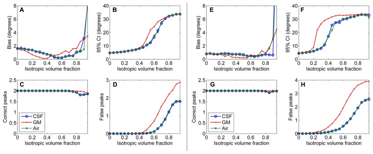
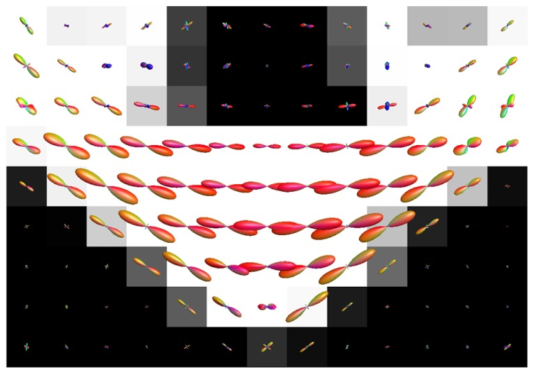
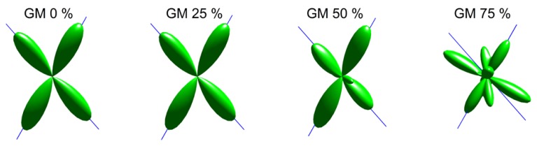
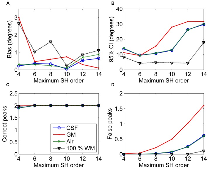
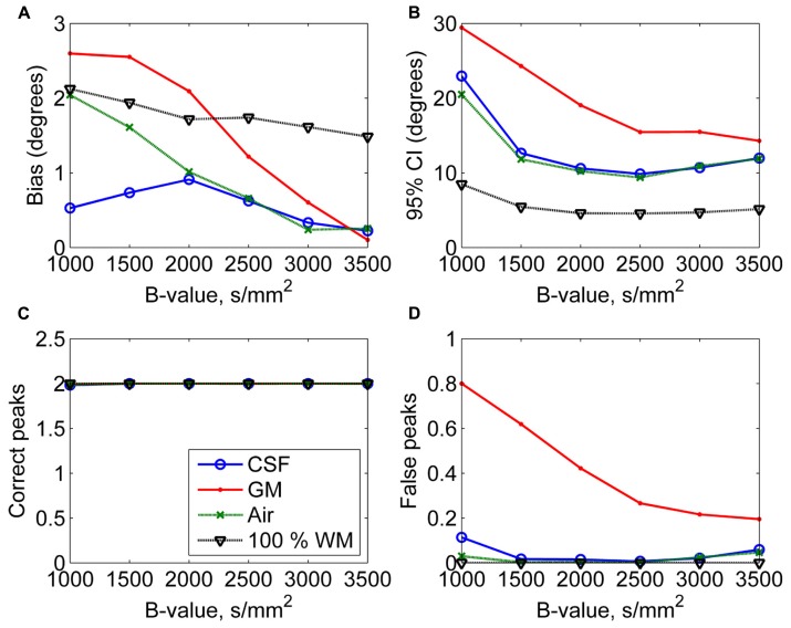
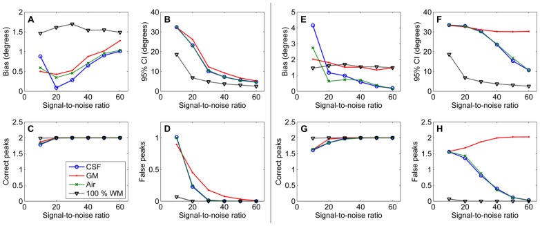
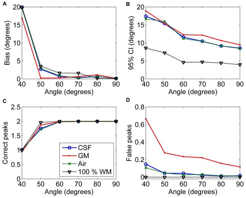
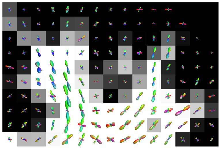
Diffusion-weighted (DW) magnetic resonance imaging (MRI) is a non-invasive imaging method, which can be used to investigate neural tracts in the white matter (WM) of the brain. Significant partial volume effects (PVEs) are present in the DW signal due to relatively large voxel sizes. These PVEs can be caused by both non-WM tissue, such as gray matter (GM) and cerebrospinal fluid (CSF), and by multiple non-parallel WM fiber populations. High angular resolution diffusion imaging (HARDI) methods have been developed to correctly characterize complex WM fiber configurations, but to date, many of the HARDI methods do not account for non-WM PVEs. In this work, we investigated the isotropic PVEs caused by non-WM tissue in WM voxels on fiber orientations extracted with constrained spherical deconvolution (CSD). Experiments were performed on simulated and real DW-MRI data. In particular, simulations were performed to demonstrate the effects of varying the diffusion weightings, signal-to-noise ratios (SNRs), fiber configurations, and tissue fractions. Our results show that the presence of non-WM tissue signal causes a decrease in the precision of the detected fiber orientations and an increase in the detection of false peaks in CSD. We estimated 35-50% of WM voxels to be affected by non-WM PVEs. For HARDI sequences, which typically have a relatively high degree of diffusion weighting, these adverse effects are most pronounced in voxels with GM PVEs. The non-WM PVEs become severe with 50% GM volume for maximum spherical harmonics orders of 8 and below, and already with 25% GM volume for higher orders. In addition, a low diffusion weighting or SNR increases the effects. The non-WM PVEs may cause problems in connectomics, where reliable fiber tracking at the WM-GM interface is especially important. We suggest acquiring data with high diffusion-weighting 2500-3000 s/mm(2), reasonable SNR (~30) and using lower SH orders in GM contaminated regions to minimize the non-WM PVEs in CSD.
弥散加权(DW)磁共振成像(MRI)是一种非侵入性成像方法,可用于研究大脑白质(WM)中的神经束。由于体素尺寸较大,DW 信号中存在显著的部分容积效应(PVE)。这些 PVE 可由非 WM 组织(如灰质(GM)和脑脊液(CSF))和多个非平行 WM 纤维群引起。高角分辨率扩散成像(HARDI)方法已被开发用于正确描述复杂的 WM 纤维结构,但迄今为止,许多 HARDI 方法并未考虑非 WM PVE。在这项工作中,我们研究了在受约束球谐分解(CSD)提取的纤维方向上,WM 体素中非 WM 组织引起的各向同性 PVE。在模拟和真实 DW-MRI 数据上进行了实验。特别是,进行了模拟以演示扩散权重、信噪比(SNR)、纤维结构和组织分数变化的影响。我们的结果表明,非 WM 组织信号的存在会降低检测到的纤维方向的精度,并增加 CSD 中虚假峰的检测。我们估计有 35-50%的 WM 体素受到非 WM PVE 的影响。对于 HARDI 序列,其通常具有相对较高的扩散加权程度,这些不利影响在具有 GM PVE 的体素中最为明显。当最大球谐阶数为 8 及以下时,GM 体积为 50%,当阶数较高时,GM 体积为 25%时,非 WM PVE 会变得严重。此外,低扩散加权或 SNR 会增加影响。非 WM PVE 可能会在连接组学中引起问题,在连接组学中,WM-GM 界面处可靠的纤维追踪尤为重要。我们建议在 GM 污染区域获取具有高扩散加权(2500-3000 s/mm2)、合理 SNR(~30)和较低 SH 阶数的数据,以最大限度地减少 CSD 中的非 WM PVE。