Gupta Manoj, Choudhury Partha S, Hazarika Dibyamohan, Rawal Sudhir
Department of Nuclear Medicine, Rajiv Gandhi Cancer Institute and Research Centre, New Delhi, India.
Department of Radiology, Rajiv Gandhi Cancer Institute and Research Centre, New Delhi, India.
World J Nucl Med. 2017 Jul-Sep;16(3):186-191. doi: 10.4103/1450-1147.207272.
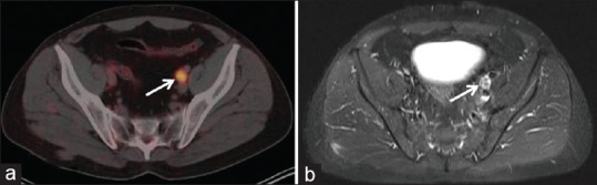
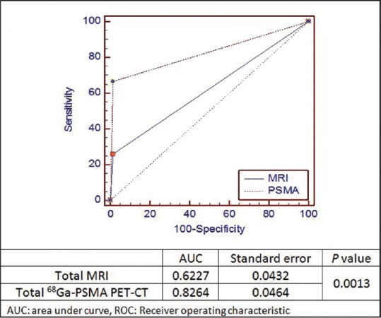
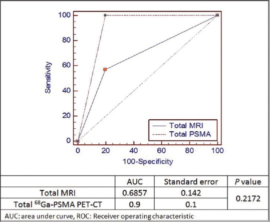
Lymph node staging plays an important role in planning initial management in nonmetastatic prostate cancer. This article compares the role of Gallium (Ga)-prostate specific membrane antigen (PSMA) positron emission tomography-computed tomography (PET-CT) with magnetic resonance imaging (MRI), which is considered the standard staging modality. Out of 39 high-risk prostate cancer patients who underwent Ga-PSMA PET-CT for staging (December 2014-December 2015), 12 patients underwent radical prostatectomy along with ePLND and were included in the analysis. Findings of the PSMA PET and MRI were compared with final histopathology. Sensitivity, specificity, positive predicative value (PPV), negative predicative value (NPV), and accuracy of Ga-PSMA PET-CT and MRI were calculated for numbers of patients and pelvic lymph node metastasis. Chi-square test, McNemar's test, and receiver operating characteristic (ROC) analysis were also done. Ga-PSMA PET-CT and MRI sensitivity, specificity, PPV, NPV, and accuracy for number of patients detection were 100%, 80%, 87.5%, 100%, 91.67%, and 57.14%, 80%, 80%, 57.4%, 66.67%, respectively. For detection of metastatic lymph node, it was 66.67%, 98.61%, 85.71%, 95.95%, 95.06% and 25.93%, 98.61%, 70%, 91.42%, 90.53%, respectively. Difference of lymph nodal detectability was statistically significant on Chi-square test. On McNemar's test, value was statistically insignificant for number of patient detection ( = 0.250) but statistically significant for lymph nodal detection ( = 0.001) for Ga-PSMA PET-CT. In ROC analysis, area under the curve was also significantly high for lymph node detectability by Ga-PSMA PET-CT. Our initial experience shows that GaPSMA PET-CT is a very promising tracer for N staging in the initial workup of prostate cancer. It has the potential to impact patient's initial management and can up- and down-stage effectively.
淋巴结分期在非转移性前列腺癌的初始治疗规划中起着重要作用。本文比较了镓(Ga)-前列腺特异性膜抗原(PSMA)正电子发射断层扫描-计算机断层扫描(PET-CT)与磁共振成像(MRI)的作用,MRI被认为是标准的分期方式。在39例接受Ga-PSMA PET-CT分期的高危前列腺癌患者(2014年12月至2015年12月)中,12例患者接受了根治性前列腺切除术及扩大盆腔淋巴结清扫术,并纳入分析。将PSMA PET和MRI的结果与最终组织病理学结果进行比较。计算Ga-PSMA PET-CT和MRI对患者数量及盆腔淋巴结转移的敏感性、特异性、阳性预测值(PPV)、阴性预测值(NPV)和准确性。还进行了卡方检验、McNemar检验和受试者工作特征(ROC)分析。Ga-PSMA PET-CT和MRI对患者数量检测的敏感性、特异性、PPV、NPV和准确性分别为100%、80%、87.5%、100%、91.67%和57.14%、80%、80%、57.4%、66.67%。对于转移性淋巴结检测,分别为66.67%、98.61%、85.71%、95.95%、95.06%和25.93%、98.61%、70%、91.42%、90.53%。卡方检验显示淋巴结可检测性差异具有统计学意义。在McNemar检验中,Ga-PSMA PET-CT对患者数量检测的P值无统计学意义(P = 0.250),但对淋巴结检测具有统计学意义(P = 0.001)。在ROC分析中,Ga-PSMA PET-CT对淋巴结可检测性的曲线下面积也显著较高。我们的初步经验表明,Ga-PSMA PET-CT在前列腺癌的初始检查中是一种非常有前景的N分期示踪剂。它有可能影响患者的初始治疗,并能有效地进行分期上调和下调。