• 文献检索
  • 文档翻译
  • 深度研究
  • 学术资讯
  • Suppr Zotero 插件Zotero 插件
  • 邀请有礼
  • 套餐&价格
  • 历史记录
应用&插件
Suppr Zotero 插件Zotero 插件浏览器插件Mac 客户端Windows 客户端微信小程序
定价
高级版会员购买积分包购买API积分包
服务
文献检索文档翻译深度研究API 文档MCP 服务
关于我们
关于 Suppr公司介绍联系我们用户协议隐私条款
关注我们

Suppr 超能文献

核心技术专利:CN118964589B侵权必究
粤ICP备2023148730 号-1Suppr @ 2026

文献检索

告别复杂PubMed语法,用中文像聊天一样搜索,搜遍4000万医学文献。AI智能推荐,让科研检索更轻松。

立即免费搜索

文件翻译

保留排版,准确专业,支持PDF/Word/PPT等文件格式,支持 12+语言互译。

免费翻译文档

深度研究

AI帮你快速写综述,25分钟生成高质量综述,智能提取关键信息,辅助科研写作。

立即免费体验

发育中牙釉质的晶体起始结构:对牙釉质晶体龋溶解的可能影响。

Crystal Initiation Structures in Developing Enamel: Possible Implications for Caries Dissolution of Enamel Crystals.

作者信息

Robinson Colin, Connell Simon D

机构信息

School of Dentistry, University of LeedsLeeds, United Kingdom.

Molecular and Nanoscale Physics Group, School of Physics and Astronomy, University of LeedsLeeds, United Kingdom.

出版信息

Front Physiol. 2017 Jun 16;8:405. doi: 10.3389/fphys.2017.00405. eCollection 2017.

DOI:10.3389/fphys.2017.00405
PMID:28670283
原文链接:https://pmc.ncbi.nlm.nih.gov/articles/PMC5472850/
Abstract

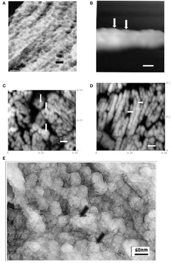
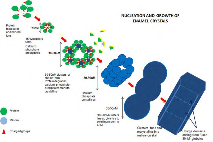
Investigations of developing enamel crystals using Atomic and Chemical Force Microscopy (AFM, CFM) have revealed a subunit structure. Subunits were seen in height images as collinear swellings about 30 nM in diameter on crystal surfaces. In friction mode they were visible as positive regions. These were similar in size (30-50 nM) to collinear spherical structures, presumably mineral matrix complexes, seen in developing enamel using a freeze fracturing/freeze etching procedure. More detailed AFM studies on mature enamel suggested that the 30-50 nM structures were composed of smaller units, ~10-15 nM in diameter. These were clustered in hexagonal or perhaps a spiral arrangement. It was suggested that these could be the imprints of initiation sites for mineral precipitation. The investigation aimed at examining original freeze etched images at high resolution to see if the smaller subunits observed using AFM in mature enamel were also present in developing enamel i.e., before loss of the organic matrix. The method used was freeze etching. Briefly samples of developing rat enamel were rapidly frozen, fractured under vacuum, and ice sublimed from the fractured surface. The fractured surface was shadowed with platinum or gold and the metal replica subjected to high resolution TEM. For AFM studies high-resolution tapping mode imaging of human mature enamel sections was performed in air under ambient conditions at a point midway between the cusp and the cervical margin. Both AFM and freeze etch studies showed structures 30-50 nM in diameter. AFM indicated that these may be clusters of somewhat smaller structures ~10-15 nM maybe hexagonally or spirally arranged. High resolution freeze etching images of very early enamel showed ~30-50 nM spherical structures in a disordered arrangement. No smaller units at 10-15 nM were clearly seen. However, when linear arrangements of 30-50 nM units were visible the picture was more complex but also smaller units including ~10-15 nM units could be observed. Structures ~10-15 nM in diameter were detected in developing enamel. While the appearance was complex, these were most evident when the 30-5 nM structures were in linear arrays. Formation of linear arrays of subunits may be associated with the development of mineral initiation sites and attendant processing of matrix proteins.

摘要

利用原子力显微镜和化学力显微镜(AFM、CFM)对发育中的釉质晶体进行的研究揭示了一种亚基结构。在高度图像中,亚基表现为晶体表面直径约30纳米的共线隆起。在摩擦模式下,它们表现为阳性区域。这些区域的大小(30 - 50纳米)与通过冷冻断裂/冷冻蚀刻程序在发育中的釉质中看到的共线球形结构(可能是矿物基质复合物)相似。对成熟釉质进行的更详细的AFM研究表明,30 - 50纳米的结构由直径约10 - 15纳米的较小单元组成。这些单元呈六边形或可能呈螺旋状排列。有人认为这些可能是矿物沉淀起始位点的印记。该研究旨在以高分辨率检查原始冷冻蚀刻图像,以确定在成熟釉质中使用AFM观察到的较小亚基在发育中的釉质(即有机基质消失之前)中是否也存在。使用的方法是冷冻蚀刻。简要来说,将发育中的大鼠釉质样本快速冷冻,在真空中断裂,然后从断裂表面升华冰。用铂或金对断裂表面进行阴影处理,并对金属复制品进行高分辨率透射电子显微镜检查。对于AFM研究,在环境条件下于空气中在牙尖和颈缘之间的中点对人类成熟釉质切片进行高分辨率轻敲模式成像。AFM和冷冻蚀刻研究均显示出直径为30 - 50纳米的结构。AFM表明这些可能是由直径约10 - 15纳米的稍小结构组成的簇,可能呈六边形或螺旋状排列。非常早期釉质的高分辨率冷冻蚀刻图像显示出直径约30 - 50纳米的球形结构呈无序排列。未清晰看到10 - 15纳米的较小单元。然而,当可见30 - 50纳米单元的线性排列时,情况更为复杂,但也能观察到包括约10 - 15纳米单元在内的较小单元。在发育中的釉质中检测到了直径约10 - 15纳米的结构。虽然其外观复杂,但当30 - 5纳米的结构呈线性阵列时最为明显。亚基线性阵列的形成可能与矿物起始位点的发育以及基质蛋白的伴随加工有关。

https://cdn.ncbi.nlm.nih.gov/pmc/blobs/8c89/5472850/ebf5e0c640e3/fphys-08-00405-g0002.jpg
https://cdn.ncbi.nlm.nih.gov/pmc/blobs/8c89/5472850/aca31464dded/fphys-08-00405-g0001.jpg
https://cdn.ncbi.nlm.nih.gov/pmc/blobs/8c89/5472850/ebf5e0c640e3/fphys-08-00405-g0002.jpg
https://cdn.ncbi.nlm.nih.gov/pmc/blobs/8c89/5472850/aca31464dded/fphys-08-00405-g0001.jpg
https://cdn.ncbi.nlm.nih.gov/pmc/blobs/8c89/5472850/ebf5e0c640e3/fphys-08-00405-g0002.jpg

相似文献

1
Crystal Initiation Structures in Developing Enamel: Possible Implications for Caries Dissolution of Enamel Crystals.发育中牙釉质的晶体起始结构:对牙釉质晶体龋溶解的可能影响。
Front Physiol. 2017 Jun 16;8:405. doi: 10.3389/fphys.2017.00405. eCollection 2017.
2
Subunit structures in hydroxyapatite crystal development in enamel: implications for amelogenesis imperfecta.釉质中羟基磷灰石晶体发育的亚基结构:对牙釉质发育不全的影响。
Connect Tissue Res. 2003;44 Suppl 1:65-71.
3
The effects of fluoride on the nanostructure and surface pK of enamel crystals: an atomic force microscopy study of human and rat enamel.氟化物对牙釉质晶体纳米结构和表面pK的影响:一项关于人类和大鼠牙釉质的原子力显微镜研究
Eur J Oral Sci. 2006 May;114 Suppl 1:99-104; discussion 127-9, 380. doi: 10.1111/j.1600-0722.2006.00275.x.
4
High resolution SE-I SEM study of enamel crystal morphology.牙釉质晶体形态的高分辨率扫描电子显微镜(二次电子成像)研究
J Electron Microsc Tech. 1990 Jan;14(1):70-8. doi: 10.1002/jemt.1060140111.
5
Two-dimensional crystals of a membrane protein: arrangement of subunits within the crystal sheet.一种膜蛋白的二维晶体:晶片中亚基的排列
Eur J Cell Biol. 1985 Mar;36(2):247-55.
6
Correlated atomic force and transmission electron microscopy of nanotubular structures in pulmonary surfactant.肺表面活性剂中纳米管状结构的相关原子力显微镜和透射电子显微镜研究
J Struct Biol. 1999 Jun 1;126(1):1-15. doi: 10.1006/jsbi.1999.4089.
7
Atomic force microscopy of freeze-fracture replicas of rat atrial tissue.大鼠心房组织冷冻断裂复制品的原子力显微镜检查。
J Microsc. 1994 Mar;173(Pt 3):173-81. doi: 10.1111/j.1365-2818.1994.tb03440.x.
8
Fine structures of cell envelopes of Chlamydia organisms as revealed by freeze-etching and negative staining techniques.通过冷冻蚀刻和负染色技术揭示的衣原体生物体细胞包膜的精细结构。
J Bacteriol. 1973 Dec;116(3):1355-63. doi: 10.1128/jb.116.3.1355-1363.1973.
9
Particle-Attachment-Mediated and Matrix/Lattice-Guided Enamel Apatite Crystal Growth.颗粒附着介导和基质/晶格导向的釉质磷灰石晶体生长。
ACS Nano. 2019 Mar 26;13(3):3151-3161. doi: 10.1021/acsnano.8b08668. Epub 2019 Feb 25.
10
In situ observation of freeze-fractured and deep-etched red blood cells with a high-vacuum low-temperature atomic force microscope.使用高真空低温原子力显微镜对冷冻断裂和深度蚀刻的红细胞进行原位观察。
Ultramicroscopy. 2005 Jan;102(2):107-14. doi: 10.1016/j.ultramic.2004.09.002.

引用本文的文献

1
Evaluating the effects of remineralizing agents on initial carious lesions.评估再矿化剂对早期龋损的影响。
Can J Dent Hyg. 2025 Feb 1;59(1):18-28. eCollection 2025 Feb.
2
Synchrotron X-ray Studies of the Structural and Functional Hierarchies in Mineralised Human Dental Enamel: A State-of-the-Art Review.矿化人类牙釉质结构与功能层次的同步加速器X射线研究:最新综述
Dent J (Basel). 2023 Apr 7;11(4):98. doi: 10.3390/dj11040098.
3
Amelogenin phosphorylation regulates tooth enamel formation by stabilizing a transient amorphous mineral precursor.

本文引用的文献

1
Biophysical characterization of synthetic amelogenin C-terminal peptides.合成牙釉蛋白C末端肽的生物物理特性
Eur J Oral Sci. 2012 Apr;120(2):113-22. doi: 10.1111/j.1600-0722.2012.00941.x. Epub 2012 Feb 11.
2
Regulation of calcium phosphate formation by amelogenins under physiological conditions.生理条件下釉原蛋白对磷酸钙形成的调控
Eur J Oral Sci. 2011 Dec;119 Suppl 1(Suppl 1):103-11. doi: 10.1111/j.1600-0722.2011.00911.x.
3
Hierarchical self-assembly of amelogenin and the regulation of biomineralization at the nanoscale.
釉原蛋白磷酸化通过稳定瞬时非晶态矿物前体来调节牙釉质形成。
J Biol Chem. 2020 Feb 14;295(7):1943-1959. doi: 10.1074/jbc.RA119.010506. Epub 2020 Jan 9.
4
The hidden structure of human enamel.人类牙釉质的隐藏结构。
Nat Commun. 2019 Sep 26;10(1):4383. doi: 10.1038/s41467-019-12185-7.
纳米级牙釉蛋白的分级自组装与生物矿化的调控。
Proc Natl Acad Sci U S A. 2011 Aug 23;108(34):14097-102. doi: 10.1073/pnas.1106228108. Epub 2011 Aug 8.
4
Transient amorphous calcium phosphate in forming enamel.正在形成的牙釉质中的瞬态无定形磷酸钙。
J Struct Biol. 2009 May;166(2):133-43. doi: 10.1016/j.jsb.2009.02.001. Epub 2009 Feb 13.
5
The effects of fluoride on the nanostructure and surface pK of enamel crystals: an atomic force microscopy study of human and rat enamel.氟化物对牙釉质晶体纳米结构和表面pK的影响:一项关于人类和大鼠牙釉质的原子力显微镜研究
Eur J Oral Sci. 2006 May;114 Suppl 1:99-104; discussion 127-9, 380. doi: 10.1111/j.1600-0722.2006.00275.x.
6
High-resolution electron microscopy of enamel-crystal demineralization and remineralization in carious lesions.龋损中釉质晶体脱矿与再矿化的高分辨率电子显微镜观察
J Electron Microsc (Tokyo). 2003;52(6):605-13. doi: 10.1093/jmicro/52.6.605.
7
Crystal growth in rat enamel.大鼠牙釉质中的晶体生长
J Cell Biol. 1963 Jul;18(1):109-23. doi: 10.1083/jcb.18.1.109.
8
Evidence for charge domains on developing enamel crystal surfaces.发育中釉质晶体表面电荷域的证据。
J Dent Res. 2000 Dec;79(12):1943-7. doi: 10.1177/00220345000790120401.
9
Effect of experimental fluorosis on the surface topography of developing enamel crystals.实验性氟中毒对正在发育的釉质晶体表面形貌的影响。
Caries Res. 2001 Jan-Feb;35(1):50-6. doi: 10.1159/000047431.
10
Subunit compartments of secretory stage enamel matrix.分泌期釉基质的亚单位区室
Connect Tissue Res. 1998;38(1-4):101-11; discussion 139-45. doi: 10.3109/03008209809017026.