• 文献检索
  • 文档翻译
  • 深度研究
  • 学术资讯
  • Suppr Zotero 插件Zotero 插件
  • 邀请有礼
  • 套餐&价格
  • 历史记录
应用&插件
Suppr Zotero 插件Zotero 插件浏览器插件Mac 客户端Windows 客户端微信小程序
定价
高级版会员购买积分包购买API积分包
服务
文献检索文档翻译深度研究API 文档MCP 服务
关于我们
关于 Suppr公司介绍联系我们用户协议隐私条款
关注我们

Suppr 超能文献

核心技术专利:CN118964589B侵权必究
粤ICP备2023148730 号-1Suppr @ 2026

文献检索

告别复杂PubMed语法,用中文像聊天一样搜索,搜遍4000万医学文献。AI智能推荐,让科研检索更轻松。

立即免费搜索

文件翻译

保留排版,准确专业,支持PDF/Word/PPT等文件格式,支持 12+语言互译。

免费翻译文档

深度研究

AI帮你快速写综述,25分钟生成高质量综述,智能提取关键信息,辅助科研写作。

立即免费体验

通过活体成像对人干细胞来源的视网膜类器官进行结构和功能表征

Structural and Functional Characterization of Human Stem-Cell-Derived Retinal Organoids by Live Imaging.

作者信息

Browne Andrew W, Arnesano Cosimo, Harutyunyan Narine, Khuu Thien, Martinez Juan Carlos, Pollack Harvey A, Koos David S, Lee Thomas C, Fraser Scott E, Moats Rex A, Aparicio Jennifer G, Cobrinik David

机构信息

USC Roski Eye Institute, Department of Ophthalmology, Keck School of Medicine of the University of Southern California, Los Angeles, California, United States.

Translational Imaging Center, University of Southern California, Los Angeles, California, United States 3Department of Molecular and Computational Biology, University of Southern California, Los Angeles, California, United States.

出版信息

Invest Ophthalmol Vis Sci. 2017 Jul 1;58(9):3311-3318. doi: 10.1167/iovs.16-20796.

DOI:10.1167/iovs.16-20796
PMID:28672397
原文链接:https://pmc.ncbi.nlm.nih.gov/articles/PMC5495152/
Abstract

PURPOSE

Human pluripotent stem cell (hPSC)-derived retinal organoids are a platform for investigating retinal development, pathophysiology, and cellular therapies. In contrast to histologic analysis in which multiple specimens fixed at different times are used to reconstruct developmental processes, repeated analysis of the same living organoids provides a more direct means to characterize changes. New live imaging modalities can provide insights into retinal organoid structure and metabolic function during in vitro growth. This study employed live tissue imaging to characterize retinal organoid development, including metabolic changes accompanying photoreceptor differentiation.

METHODS

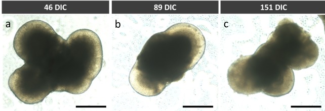
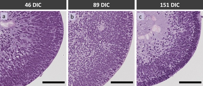
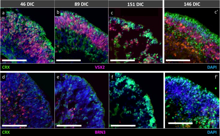
Live hPSC-derived retinal organoids at different developmental stages were examined for microanatomic organization and metabolic function by phase contrast microscopy, optical coherence tomography (OCT), fluorescence lifetime imaging microscopy (FLIM), and hyperspectral imaging (HSpec). Features were compared to those revealed by histologic staining, immunostaining, and microcomputed tomography (micro-CT) of fixed organoid tissue.

RESULTS

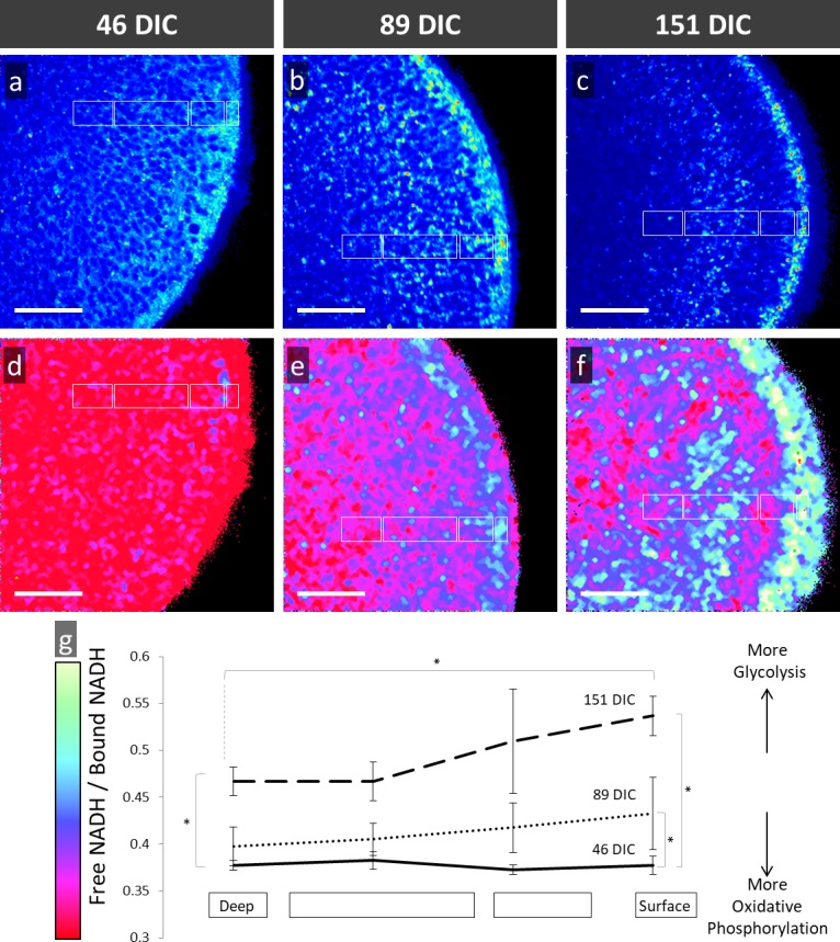
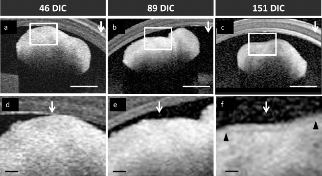
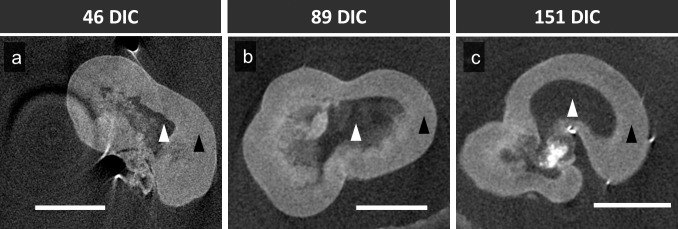
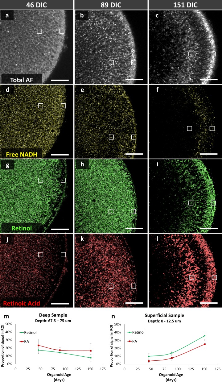
We used FLIM and HSpec to detect changes in metabolic activity as organoids differentiated into organized lamellae. FLIM detected increased glycolytic activity and HSpec detected retinol and retinoic acid accumulation in the organoid outer layer, coinciding with photoreceptor genesis. OCT enabled imaging of lamellae formed during organoid maturation. Micro-CT revealed three-dimensional structure, but failed to detect lamellae.

CONCLUSIONS

Live imaging modalities facilitate real-time and nondestructive imaging of retinal organoids as they organize into lamellar structures. FLIM and HSpec enable rapid detection of lamellar structure and photoreceptor metabolism. Live imaging techniques may aid in the continuous evaluation of retinal organoid development in diverse experimental and cell therapy settings.

摘要

目的

人多能干细胞(hPSC)来源的视网膜类器官是研究视网膜发育、病理生理学和细胞治疗的一个平台。与使用在不同时间固定的多个标本重建发育过程的组织学分析不同,对同一活类器官进行重复分析提供了一种更直接的方法来表征变化。新的活体成像方式可以深入了解体外生长过程中视网膜类器官的结构和代谢功能。本研究采用活体组织成像来表征视网膜类器官的发育,包括伴随光感受器分化的代谢变化。

方法

通过相差显微镜、光学相干断层扫描(OCT)、荧光寿命成像显微镜(FLIM)和高光谱成像(HSpec),对不同发育阶段的活hPSC来源的视网膜类器官进行微观解剖组织和代谢功能检查。将这些特征与固定类器官组织的组织学染色、免疫染色和微型计算机断层扫描(micro-CT)所显示的特征进行比较。

结果

我们使用FLIM和HSpec来检测类器官分化为有组织的板层时代谢活性的变化。FLIM检测到糖酵解活性增加,HSpec检测到类器官外层视黄醇和视黄酸的积累,这与光感受器的发生相吻合。OCT能够对类器官成熟过程中形成的板层进行成像。Micro-CT揭示了三维结构,但未能检测到板层。

结论

活体成像方式有助于对视网膜类器官组织成板层结构的过程进行实时和非破坏性成像。FLIM和HSpec能够快速检测板层结构和光感受器代谢。活体成像技术可能有助于在各种实验和细胞治疗环境中对视网膜类器官的发育进行持续评估。

https://cdn.ncbi.nlm.nih.gov/pmc/blobs/ef21/5495152/506349006334/i1552-5783-58-9-3311-f07.jpg
https://cdn.ncbi.nlm.nih.gov/pmc/blobs/ef21/5495152/c8472a06bb45/i1552-5783-58-9-3311-f01.jpg
https://cdn.ncbi.nlm.nih.gov/pmc/blobs/ef21/5495152/5411e9b40fdc/i1552-5783-58-9-3311-f02.jpg
https://cdn.ncbi.nlm.nih.gov/pmc/blobs/ef21/5495152/92a2da775e95/i1552-5783-58-9-3311-f03.jpg
https://cdn.ncbi.nlm.nih.gov/pmc/blobs/ef21/5495152/f42abc57379c/i1552-5783-58-9-3311-f04.jpg
https://cdn.ncbi.nlm.nih.gov/pmc/blobs/ef21/5495152/41a62fe11f97/i1552-5783-58-9-3311-f05.jpg
https://cdn.ncbi.nlm.nih.gov/pmc/blobs/ef21/5495152/c88f39698735/i1552-5783-58-9-3311-f06.jpg
https://cdn.ncbi.nlm.nih.gov/pmc/blobs/ef21/5495152/506349006334/i1552-5783-58-9-3311-f07.jpg

相似文献

1
Structural and Functional Characterization of Human Stem-Cell-Derived Retinal Organoids by Live Imaging.通过活体成像对人干细胞来源的视网膜类器官进行结构和功能表征
Invest Ophthalmol Vis Sci. 2017 Jul 1;58(9):3311-3318. doi: 10.1167/iovs.16-20796.
2
Retinal Organoids Long-Term Functional Characterization Using Two-Photon Fluorescence Lifetime and Hyperspectral Microscopy.使用双光子荧光寿命和高光谱显微镜对视网膜类器官进行长期功能表征
Front Cell Neurosci. 2021 Dec 10;15:796903. doi: 10.3389/fncel.2021.796903. eCollection 2021.
3
Reproducibility and staging of 3D human retinal organoids across multiple pluripotent stem cell lines.在多个多能干细胞系中重现和分期 3D 人视网膜类器官。
Development. 2019 Jan 9;146(1):dev171686. doi: 10.1242/dev.171686.
4
Three-Dimensional Retinal Organoids Facilitate the Investigation of Retinal Ganglion Cell Development, Organization and Neurite Outgrowth from Human Pluripotent Stem Cells.三维视网膜类器官促进人多能干细胞中视网膜神经节细胞发育、组织和神经突生长的研究。
Sci Rep. 2018 Sep 28;8(1):14520. doi: 10.1038/s41598-018-32871-8.
5
Transplanted hESC-Derived Retina Organoid Sheets Differentiate, Integrate, and Improve Visual Function in Retinal Degenerate Rats.移植的 hESC 衍生视网膜类器官片在视网膜变性大鼠中分化、整合并改善视觉功能。
Invest Ophthalmol Vis Sci. 2018 May 1;59(6):2586-2603. doi: 10.1167/iovs.17-23646.
6
Differentiation of retinal organoids from human pluripotent stem cells.人多能干细胞来源的视网膜类器官的分化。
Methods Cell Biol. 2020;159:279-302. doi: 10.1016/bs.mcb.2020.02.005. Epub 2020 Mar 11.
7
Organoid technology for retinal repair.用于视网膜修复的类器官技术。
Dev Biol. 2018 Jan 15;433(2):132-143. doi: 10.1016/j.ydbio.2017.09.028. Epub 2017 Dec 25.
8
Rapid and efficient generation of mature retinal organoids derived from human pluripotent stem cells via optimized pharmacological modulation of Sonic hedgehog, activin A, and retinoic acid signal transduction.通过优化 Sonic hedgehog、激活素 A 和视黄酸信号转导的药理学调节,从人多能干细胞快速有效地生成成熟的视网膜类器官。
PLoS One. 2024 Aug 9;19(8):e0308743. doi: 10.1371/journal.pone.0308743. eCollection 2024.
9
A highly reproducible and efficient method for retinal organoid differentiation from human pluripotent stem cells.一种高度可重现且高效的方法,用于从人类多能干细胞中分化出视网膜类器官。
Proc Natl Acad Sci U S A. 2024 Jun 18;121(25):e2317285121. doi: 10.1073/pnas.2317285121. Epub 2024 Jun 13.
10
Antioxidant and lipid supplementation improve the development of photoreceptor outer segments in pluripotent stem cell-derived retinal organoids.抗氧化和脂质补充可改善多能干细胞衍生的视网膜类器官中外节感光细胞的发育。
Stem Cell Reports. 2022 Apr 12;17(4):775-788. doi: 10.1016/j.stemcr.2022.02.019. Epub 2022 Mar 24.

引用本文的文献

1
How to Study the Mechanobiology of Intestinal Epithelial Organoids? A Review of Culture Supports, Imaging Techniques, and Analysis Methods.如何研究肠道上皮类器官的力学生物学?培养支架、成像技术及分析方法综述
Biol Cell. 2025 Apr;117(4):e70003. doi: 10.1111/boc.70003.
2
Retina-specific laminin-based generation of photoreceptor progenitors from human pluripotent stem cells under xeno-free and chemically defined conditions.在无血清和化学成分明确的条件下,基于层粘连蛋白从人多能干细胞中特异性生成视网膜光感受器祖细胞。
Nat Protoc. 2025 Feb 25. doi: 10.1038/s41596-025-01142-y.
3
Single-Cell Transcriptomic Dataset of RPGR-associated Retinitis Pigmentosa Patient-Derived Retinal Organoids.

本文引用的文献

1
Hyperspectral phasor analysis enables multiplexed 5D in vivo imaging.高光谱相位分析可实现 5D 体内成像的多重化。
Nat Methods. 2017 Feb;14(2):149-152. doi: 10.1038/nmeth.4134. Epub 2017 Jan 9.
2
Temporal expression of CD184(CXCR4) and CD171(L1CAM) identifies distinct early developmental stages of human retinal ganglion cells in embryonic stem cell derived retina.CD184(CXCR4)和CD171(L1CAM)的时序表达确定了胚胎干细胞来源视网膜中人类视网膜神经节细胞不同的早期发育阶段。
Exp Eye Res. 2017 Jan;154:177-189. doi: 10.1016/j.exer.2016.11.013. Epub 2016 Nov 17.
3
Organoids as an in vitro model of human development and disease.
RPGR 相关性视网膜炎患者来源的视网膜类器官的单细胞转录组数据集。
Sci Data. 2024 Nov 26;11(1):1285. doi: 10.1038/s41597-024-04124-z.
4
Harnessing the power of artificial intelligence for human living organoid research.利用人工智能的力量进行人类类器官研究。
Bioact Mater. 2024 Aug 30;42:140-164. doi: 10.1016/j.bioactmat.2024.08.027. eCollection 2024 Dec.
5
Optical redox imaging to screen synthetic hydrogels for stem cell-derived cardiomyocyte differentiation and maturation.用于筛选促进干细胞衍生心肌细胞分化和成熟的合成水凝胶的光学氧化还原成像。
Biophotonics Discov. 2024 May;1(1). doi: 10.1117/1.bios.1.1.015002. Epub 2024 May 20.
6
A treatment within sight: challenges in the development of stem cell-derived photoreceptor therapies for retinal degenerative diseases.一种即将实现的治疗方法:视网膜退行性疾病干细胞衍生光感受器疗法开发中的挑战
Front Transplant. 2023 Sep 29;2:1130086. doi: 10.3389/frtra.2023.1130086. eCollection 2023.
7
Deep learning based characterization of human organoids using optical coherence tomography.基于深度学习的光学相干断层扫描对人类类器官的表征
Biomed Opt Express. 2024 Apr 17;15(5):3112-3127. doi: 10.1364/BOE.515781. eCollection 2024 May 1.
8
Label-free metabolic optical biomarkers track stem cell fate transition in real time.无标记代谢光学生物标志物实时跟踪干细胞命运转变。
Sci Adv. 2024 May 10;10(19):eadi6770. doi: 10.1126/sciadv.adi6770. Epub 2024 May 8.
9
Visualisation of gene expression within the context of tissues using an X-ray computed tomography-based multimodal approach.基于 X 射线计算机断层扫描的多模态方法在组织中可视化基因表达。
Sci Rep. 2024 Apr 12;14(1):8543. doi: 10.1038/s41598-024-58766-5.
10
Semiconducting polymer dots for multifunctional integrated nanomedicine carriers.用于多功能集成纳米医学载体的半导体聚合物点
Mater Today Bio. 2024 Mar 24;26:101028. doi: 10.1016/j.mtbio.2024.101028. eCollection 2024 Jun.
类器官作为人类发育和疾病的体外模型。
Nat Cell Biol. 2016 Mar;18(3):246-54. doi: 10.1038/ncb3312.
4
Concise Review: Fluorescent Reporters in Human Pluripotent Stem Cells: Contributions to Cardiac Differentiation and Their Applications in Cardiac Disease and Toxicity.简要综述:人类多能干细胞中的荧光报告基因:对心脏分化的贡献及其在心脏病和毒性研究中的应用
Stem Cells. 2016 Jan;34(1):13-26. doi: 10.1002/stem.2196. Epub 2015 Oct 8.
5
Transcriptome Dynamics of Developing Photoreceptors in Three-Dimensional Retina Cultures Recapitulates Temporal Sequence of Human Cone and Rod Differentiation Revealing Cell Surface Markers and Gene Networks.三维视网膜培养物中发育中的光感受器的转录组动态概括了人类视锥细胞和视杆细胞分化的时间序列,揭示了细胞表面标志物和基因网络。
Stem Cells. 2015 Dec;33(12):3504-18. doi: 10.1002/stem.2122. Epub 2015 Aug 14.
6
Simultaneous decomposition of multiple hyperspectral data sets: signal recovery of unknown fluorophores in the retinal pigment epithelium.多个高光谱数据集的同步分解:视网膜色素上皮中未知荧光团的信号恢复
Biomed Opt Express. 2014 Nov 6;5(12):4171-85. doi: 10.1364/BOE.5.004171. eCollection 2014 Dec 1.
7
Dynamic Assessment of the Endothelialization of Tissue-Engineered Blood Vessels Using an Optical Coherence Tomography Catheter-Based Fluorescence Imaging System.使用基于光学相干断层扫描导管的荧光成像系统对组织工程血管内皮化进行动态评估
Tissue Eng Part C Methods. 2015 Jul;21(7):758-66. doi: 10.1089/ten.TEC.2014.0345. Epub 2015 Jan 30.
8
Anatomy of the fetal membranes using optical coherence tomography: part 1.使用光学相干断层扫描对胎膜的解剖结构:第1部分。
Placenta. 2014 Dec;35(12):1065-9. doi: 10.1016/j.placenta.2014.09.011. Epub 2014 Sep 28.
9
Organogenesis in a dish: modeling development and disease using organoid technologies.器官发生在培养皿中:使用类器官技术模拟发育和疾病。
Science. 2014 Jul 18;345(6194):1247125. doi: 10.1126/science.1247125. Epub 2014 Jul 17.
10
Generation of three-dimensional retinal tissue with functional photoreceptors from human iPSCs.利用人类诱导多能干细胞生成具有功能性光感受器的三维视网膜组织。
Nat Commun. 2014 Jun 10;5:4047. doi: 10.1038/ncomms5047.