Preißler Sandra, Thielemann Désirée, Dietrich Caroline, Hofmann Gunther O, Miltner Wolfgang H R, Weiss Thomas
Department of Biological and Clinical Psychology, Friedrich Schiller UniversityJena, Germany.
Clinic for Trauma and Reconstructive Surgery, Berufsgenossenschaftliche Kliniken Bergmannstrost HalleHalle, Germany.
Front Hum Neurosci. 2017 Jun 20;11:319. doi: 10.3389/fnhum.2017.00319. eCollection 2017.
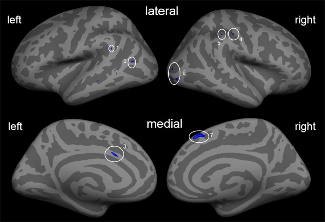
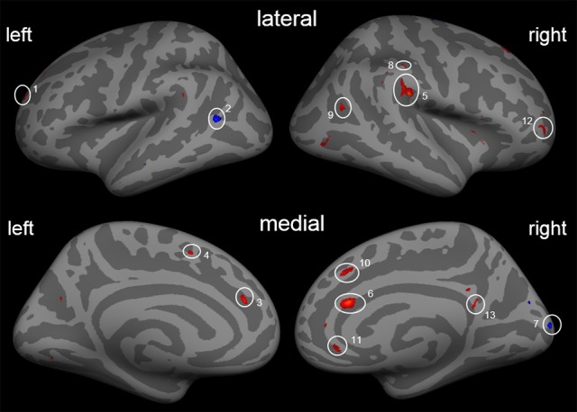
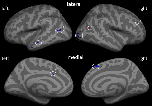
The aim of this study was to investigate whether a special prosthetic training in phantom limb pain patients aimed at increasing the functional use of the prosthesis leads to neural morphological plasticity of brain structures and a reduction in phantom limb pain. For chronic pain disorders, it was shown that morphological alterations due to pain might become at least partially reversed by pain therapies. Phantom limb pain is a chronic pain disorder that is frequently followed by neural plasticity of anatomical brain structures. In our study, 10 patients with amputation of the upper limb participated in a two-week training with a myoelectric prosthesis with somatosensory feedback. Grip strength was fed back with electrocutaneous stimulus patterns applied to the stump. Phantom limb pain was assessed before and after the two-week training. Similarly, two T1 weighted MRI scans were conducted for longitudinal thickness analyses of cortical brain structures. As result of this treatment, patients experienced a reduction in phantom limb pain and a gain in prosthesis functionality. Furthermore, we found a change of cortical thickness in small brain areas in the visual stream and the post-central gyrus ipsilateral to the amputation indicating morphological alterations in brain areas involved in vision and pain processing.
本研究的目的是调查针对幻肢痛患者的特殊假肢训练,旨在增加假肢的功能使用是否会导致脑结构的神经形态可塑性以及幻肢痛的减轻。对于慢性疼痛障碍,研究表明疼痛引起的形态改变可能至少部分地通过疼痛治疗得到逆转。幻肢痛是一种慢性疼痛障碍,常伴有解剖学脑结构的神经可塑性。在我们的研究中,10名上肢截肢患者参与了为期两周的使用带有体感反馈的肌电假肢的训练。握力通过施加于残端的电刺激模式进行反馈。在为期两周的训练前后对幻肢痛进行了评估。同样,进行了两次T1加权MRI扫描,以对脑皮质结构进行纵向厚度分析。作为这种治疗的结果,患者的幻肢痛减轻,假肢功能得到改善。此外,我们发现视觉流和截肢同侧中央后回的小脑区皮质厚度发生了变化,这表明参与视觉和疼痛处理的脑区出现了形态改变。