Paniagua Beatriz, Pascal Laura, Prieto Juan, Vimort Jean Baptiste, Gomes Liliane, Yatabe Marilia, Ruellas Antonio Carlos, Budin Francois, Pieper Steve, Styner Martin, Benavides Erika, Cevidanes Lucia
Kitware Inc., 101 Weaver St Suite G4, Carrboro, NC, USA 27510.
University of Michigan, School of Dentistry, Department of Orthodontics and Pediatric Dentistry, 1011 North University Avenue, Ann Arbor, Michigan 48109, United States.
Proc SPIE Int Soc Opt Eng. 2017 Feb 11;10137. doi: 10.1117/12.2254070. Epub 2017 Mar 13.
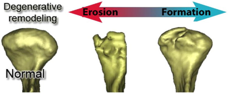
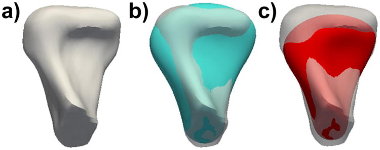
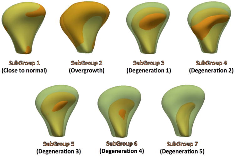
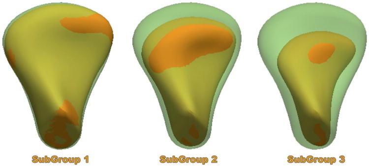
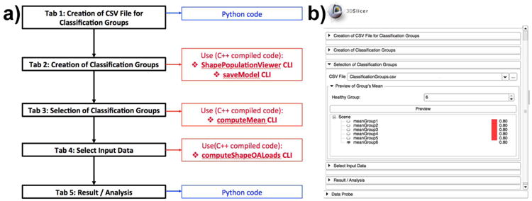
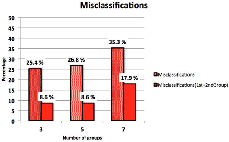
Osteoarthritis (OA) of temporomandibular joints (TMJ) occurs in about 40% of the patients who present TMJ disorders. Despite its prevalence, OA diagnosis and treatment remain controversial since there are no clear symptoms of the disease, especially in early stages. Quantitative tools based on 3D imaging of the TMJ condyle have the potential to help characterize TMJ OA changes. The goals of the tools proposed in this study are to ultimately develop robust imaging markers for diagnosis and assessment of treatment efficacy. This work proposes to identify differences among asymptomatic controls and different clinical phenotypes of TMJ OA by means of Statistical Shape Modeling (SSM), obtained via clinical expert consensus. From three different grouping schemes (with 3, 5 and 7 groups), our best results reveal that that the majority (74.5%) of the classifications occur in agreement with the groups assigned by consensus between our clinical experts. Our findings suggest the existence of different disease-based phenotypic morphologies in TMJ OA. Our preliminary findings with statistical shape modeling based biomarkers may provide a quantitative staging of the disease. The methodology used in this study is included in an open source image analysis toolbox, to ensure reproducibility and appropriate distribution and dissemination of the solution proposed.
颞下颌关节骨关节炎(OA)发生于约40%出现颞下颌关节紊乱的患者中。尽管其患病率较高,但由于该疾病没有明确的症状,尤其是在早期阶段,OA的诊断和治疗仍存在争议。基于颞下颌关节髁突三维成像的定量工具有可能帮助描述颞下颌关节OA的变化特征。本研究中提出的工具的目标是最终开发出用于诊断和评估治疗效果的可靠成像标志物。这项工作建议通过基于临床专家共识获得的统计形状模型(SSM)来识别无症状对照与颞下颌关节OA不同临床表型之间的差异。从三种不同的分组方案(3组、5组和7组)来看,我们的最佳结果显示,大多数(74.5%)分类与我们临床专家通过共识指定的组一致。我们的研究结果表明,颞下颌关节OA中存在基于疾病的不同表型形态。我们基于统计形状模型的生物标志物的初步研究结果可能为该疾病提供定量分期。本研究中使用的方法包含在一个开源图像分析工具箱中,以确保所提出解决方案的可重复性以及适当的分发和传播。