Choi Sooyoung, Noh Daji, Kim Youngwhan, Jeong Inseong, Choi Hojung, Lee Youngwon, Lee Kija
Ian Animal Diagnostic Center, Seoul 06014, Korea.
College of Veterinary Medicine, Kyungpook National University, Daegu 41566, Korea.
J Vet Sci. 2018 Jan 31;19(1):137-142. doi: 10.4142/jvs.2018.19.1.137.
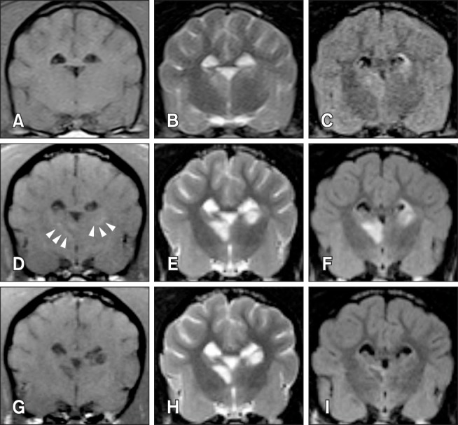
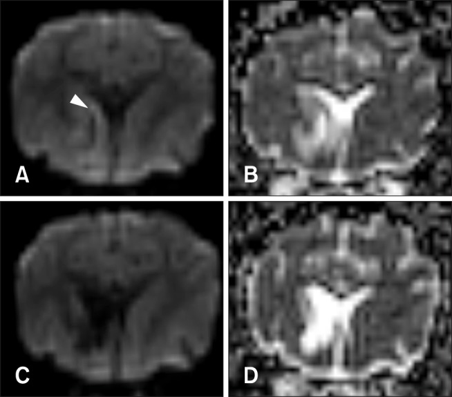
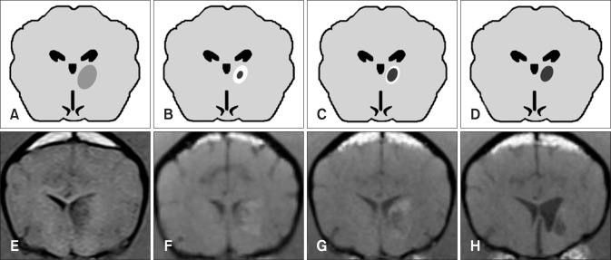
This study describes magnetic resonance imaging (MRI) results and changes in lateral ventricular size over time in a canine ischemic stroke model. T1- and T2-weighted (T1W, T2W) imaging and fluid-attenuated inversion recovery (FLAIR) sequence MRI were performed at 3 h and 3, 8, and 35 days after brain infarct induction. Diffusion-weighted imaging (DWI) and apparent diffusion coefficient (ADC) mapping were performed at 8 and 35 days. A total of 29 brain lesions were induced successfully in 12 of 14 beagle dogs. At 3 h, T2W and FLAIR detected hyperintense lesions in three randomly selected dogs. On T1W, all lesions appeared hypointense to isointense at 3 h, isointense (18/29) or hypointense (11/29) at 3 days, hypointense to isointense with peripheral hyperintensity (24/26) at 8 days, and hypointense (18/26) at 35 days. Infarcts on DWI/ADC were hypointense to isointense centrally, with the periphery hyperintense/hyperintense (17/26) at 8 days and hypointense/hyperintense (19/26) at 35 days. A marked increase in lateral ventricular size was observed in dogs with cerebral infarcts. In conclusion, T2W and FLAIR were useful for detecting early stage (3 h to 3 days) brain infarction. T1W and DWI were useful for detecting neuronal necrosis and providing supplemental information for phase evaluation.
本研究描述了犬缺血性中风模型中磁共振成像(MRI)结果以及侧脑室大小随时间的变化。在脑梗死诱导后3小时以及3、8和35天进行了T1加权(T1W)和T2加权(T2W)成像以及液体衰减反转恢复(FLAIR)序列MRI检查。在8天和35天进行了扩散加权成像(DWI)和表观扩散系数(ADC)图检查。在14只比格犬中的12只中成功诱导出了总共29个脑损伤。在3小时时,T2W和FLAIR在三只随机选择的犬中检测到高信号损伤。在T1W上,所有损伤在3小时时呈低信号至等信号,在3天时呈等信号(18/29)或低信号(11/29),在8天时呈低信号至等信号并伴有周边高信号(24/26),在35天时呈低信号(18/26)。DWI/ADC上的梗死灶中央呈低信号至等信号,周边在8天时呈高信号/高信号(17/26),在35天时呈低信号/高信号(19/26)。在患有脑梗死的犬中观察到侧脑室大小显著增加。总之,T2W和FLAIR对于检测早期(3小时至3天)脑梗死很有用。T1W和DWI对于检测神经元坏死以及为分期评估提供补充信息很有用。