Shiels Stefanie M, Talley Anne D, McGough Madison A P, Zienkiewicz Katarzyna J, Kalpakci Kerem, Shimko Daniel, Guelcher Scott A, Wenke Joseph C
US Army Institute of Surgical Research, Fort Sam Houston, TX, USA.
Department of Chemical and Biomolecular Engineering, Vanderbilt University, Nashville, TN, USA.
J Orthop Surg Res. 2017 Jul 11;12(1):107. doi: 10.1186/s13018-017-0613-0.
The challenging biological and mechanical environment of posterolateral fusion (PLF) requires a carrier that spans the transverse processes and resists the compressive forces of the posterior musculature. The less traumatic posterolateral approach enabled by minimally invasive surgical techniques has prompted investigations into alternative rhBMP-2 carriers that are injectable, settable, and compression-resistant. In this pilot study, we investigated injectable low-viscosity (LV) polymer/composite bone grafts as compression-resistant carriers for rhBMP-2 in a single-level rabbit PLF model.
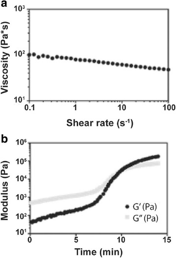
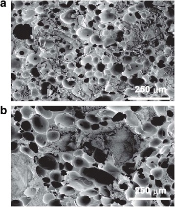
LV grafts were augmented with ceramic microparticles: (1) hydrolytically degradable bioactive glass (BG), or (2) cell-degradable 85% β-tricalcium phosphate/15% hydroxyapatite (CM). Material properties, such as pore size, viscosity, working time, and bulk modulus upon curing, were measured for each LV polymer/ceramic material. An in vivo model of posterolateral fusion in a rabbit was used to assess the grafts' capability to encourage spinal fusion.
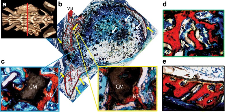
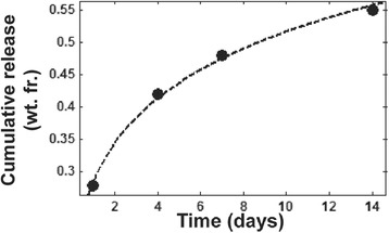
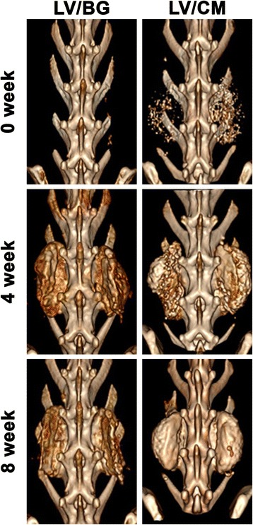
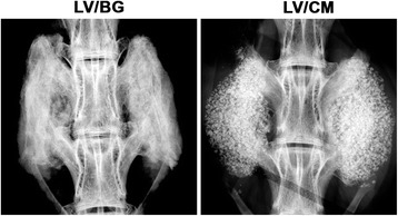
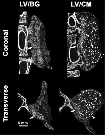
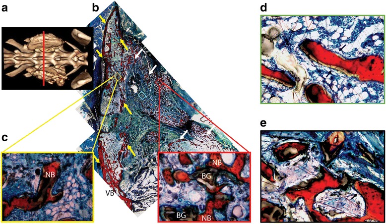
These materials maintained a working time between 9.6 and 10.3 min, with a final bulk modulus between 1.2 and 3.1 MPa. The LV polymer/composite bone grafts released 55% of their rhBMP-2 over a 14-day period. As assessed by manual palpation in vivo, fusion was achieved in all (n = 3) animals treated with LV/BG or LV/CM carriers incorporating 430 μg rhBMP-2/ml. Images of μCT and histological sections revealed evidence of bone fusion near the transverse processes.
This study highlights the potential of LV grafts as injectable and compression-resistant rhBMP-2 carriers for posterolateral spinal fusion.
后外侧融合术(PLF)面临的生物和力学环境具有挑战性,需要一种能够跨越横突并抵抗后部肌肉组织压缩力的载体。微创外科技术实现的创伤较小的后外侧入路促使人们对可注射、可固化且抗压的重组人骨形态发生蛋白-2(rhBMP-2)替代载体进行研究。在这项前瞻性研究中,我们在单节段兔PLF模型中研究了可注射低粘度(LV)聚合物/复合骨移植材料作为rhBMP-2的抗压载体。
LV移植物用陶瓷微粒增强:(1)可水解降解的生物活性玻璃(BG),或(2)细胞可降解的85%β-磷酸三钙/15%羟基磷灰石(CM)。测量每种LV聚合物/陶瓷材料的材料特性,如孔径、粘度、工作时间和固化后的体积模量。使用兔后外侧融合的体内模型评估移植物促进脊柱融合的能力。
这些材料的工作时间维持在9.6至10.3分钟之间,最终体积模量在1.2至3.1兆帕之间。LV聚合物/复合骨移植物在14天内释放了55%的rhBMP-2。通过体内手动触诊评估,所有(n = 3)接受含430μg rhBMP-2/ml的LV/BG或LV/CM载体治疗的动物均实现了融合。μCT图像和组织学切片显示横突附近有骨融合的证据。
本研究突出了LV移植物作为用于后外侧脊柱融合的可注射且抗压的rhBMP-2载体的潜力。