Rueckl Martin, Lenzi Stephen C, Moreno-Velasquez Laura, Parthier Daniel, Schmitz Dietmar, Ruediger Sten, Johenning Friedrich W
Institute of Physics, Humboldt Universität BerlinBerlin, Germany.
Neuroscience Research Center, Charité Universitätsmedizin BerlinBerlin, Germany.
Front Neuroinform. 2017 Jun 29;11:44. doi: 10.3389/fninf.2017.00044. eCollection 2017.
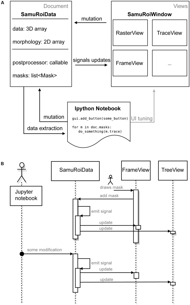
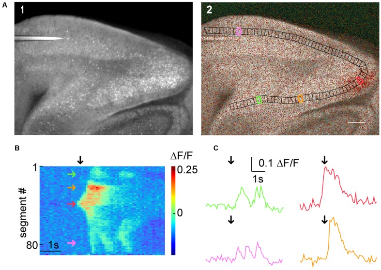
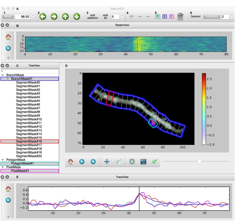
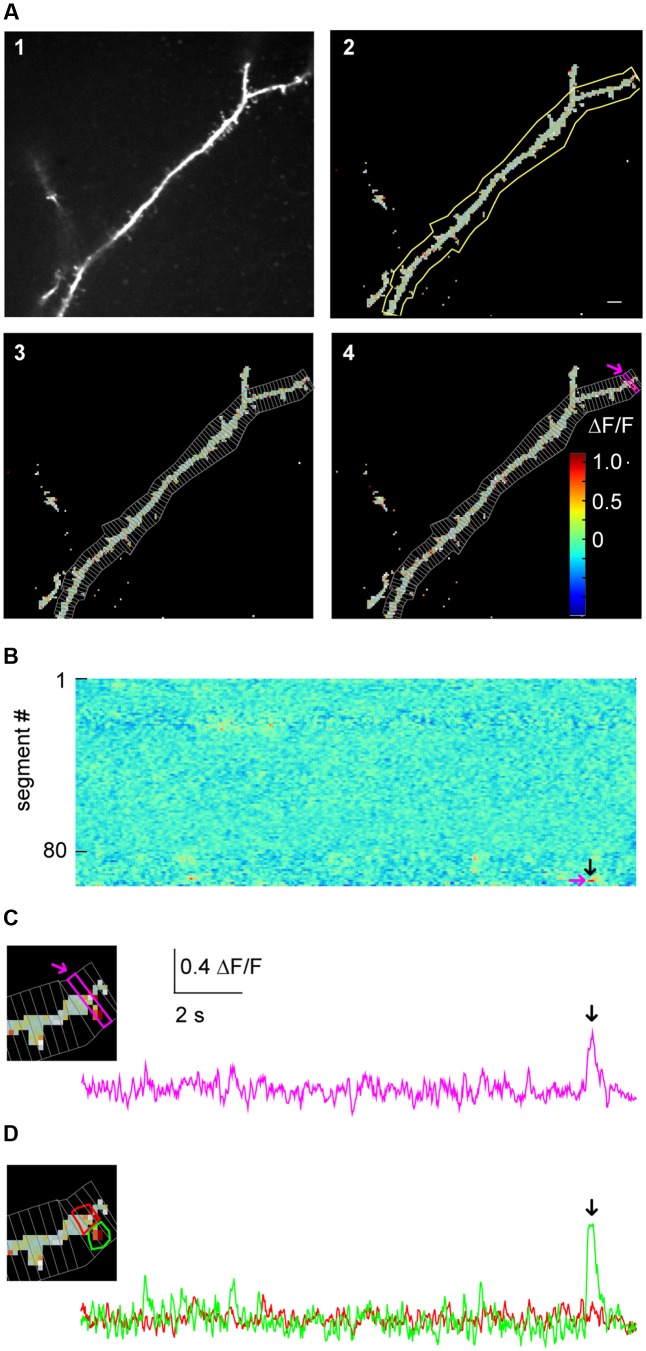
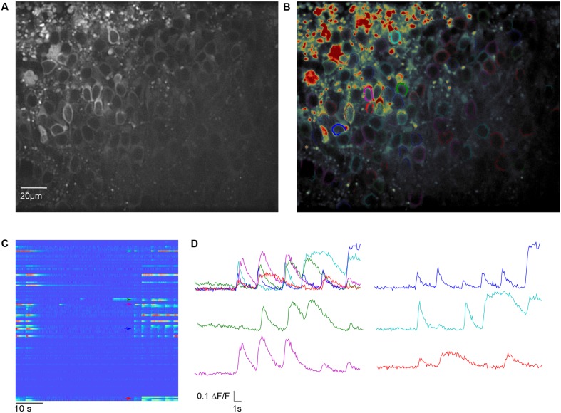
The measurement of activity and has shifted from electrical to optical methods. While the indicators for imaging activity have improved significantly over the last decade, tools for analysing optical data have not kept pace. Most available analysis tools are limited in their flexibility and applicability to datasets obtained at different spatial scales. Here, we present SamuROI (Structured analysis of multiple user-defined ROIs), an open source Python-based analysis environment for imaging data. SamuROI simplifies exploratory analysis and visualization of image series of fluorescence changes in complex structures over time and is readily applicable at different spatial scales. In this paper, we show the utility of SamuROI in Ca-imaging based applications at three spatial scales: the micro-scale (i.e., sub-cellular compartments including cell bodies, dendrites and spines); the meso-scale, (i.e., whole cell and population imaging with single-cell resolution); and the macro-scale (i.e., imaging of changes in bulk fluorescence in large brain areas, without cellular resolution). The software described here provides a graphical user interface for intuitive data exploration and region of interest (ROI) management that can be used interactively within Jupyter Notebook: a publicly available interactive Python platform that allows simple integration of our software with existing tools for automated ROI generation and post-processing, as well as custom analysis pipelines. SamuROI software, source code and installation instructions are publicly available on GitHub and documentation is available online. SamuROI reduces the energy barrier for manual exploration and semi-automated analysis of spatially complex Ca imaging datasets, particularly when these have been acquired at different spatial scales.
活动测量方式已从电学方法转变为光学方法。尽管在过去十年中成像活动指标有了显著改善,但用于分析光学数据的工具却未能跟上步伐。大多数现有的分析工具在灵活性以及对不同空间尺度获取的数据集的适用性方面都存在局限。在此,我们介绍SamuROI(多用户定义感兴趣区域的结构化分析),这是一个基于Python的用于成像数据的开源分析环境。SamuROI简化了对复杂结构中荧光随时间变化的图像序列的探索性分析和可视化,并且可轻松应用于不同空间尺度。在本文中,我们展示了SamuROI在基于钙成像的应用中在三个空间尺度上的效用:微观尺度(即亚细胞区室,包括细胞体、树突和棘突);中观尺度(即具有单细胞分辨率的全细胞和群体成像);以及宏观尺度(即对大脑大区域中整体荧光变化的成像,无细胞分辨率)。这里描述的软件提供了一个图形用户界面,用于直观的数据探索和感兴趣区域(ROI)管理,可在Jupyter Notebook中进行交互式使用:Jupyter Notebook是一个公开可用的交互式Python平台,它允许我们的软件与用于自动生成ROI和后处理的现有工具以及自定义分析管道进行简单集成。SamuROI软件、源代码和安装说明可在GitHub上公开获取,文档也可在线获取。SamuROI降低了对空间复杂的钙成像数据集进行手动探索和半自动分析的能量障碍,特别是当这些数据集是在不同空间尺度上获取时。