Shin Seung Kak, Kim Yun Soo, Choi Seung Joon, Shim Young Sup, Jung Dong Hae, Kwon Oh Sang, Choi Duck Joo, Kim Ju Hyun
Department of Internal medicine Department of Radiology Department of Pathology, Gachon University Gil Medical Center, Incheon, Republic of Korea.
Medicine (Baltimore). 2017 Jul;96(29):e7278. doi: 10.1097/MD.0000000000007278.
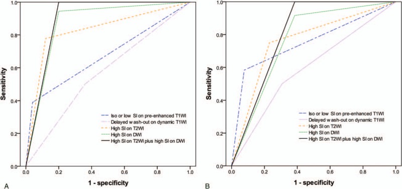
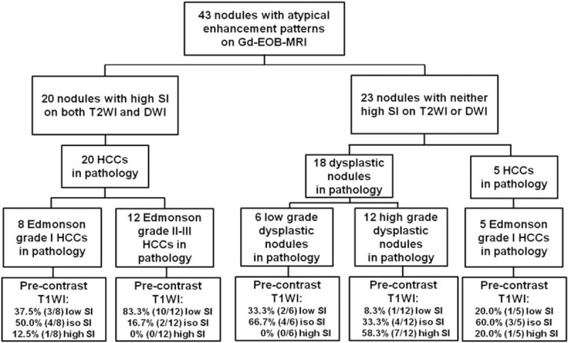
It is difficult to characterize the nodular lesions in cirrhotic liver if typical enhancement pattern is not present on dynamic contrast-enhanced imagings. Although the signal intensity of the hepatobiliary phase in gadoxetic acid-enhanced magnetic resonance imaging (MRI) is helpful for characterization of the lesions, some dysplastic nodules may also exhibit low signal intensity in the hepatobiliary phase. We aimed to assess the usefulness of gadoxetic acid (Gd-EOB-DTPA)-enhanced MRI including diffusion-weighted imaging (DWI) for differentiation between atypical small hepatocellular carcinomas (HCCs) and dysplastic nodules showing low signal intensity (SI) in the hepatobiliary phase, and to evaluate the MRI findings in determining the histological grade of atypical HCCs in patients with cirrhosis.A total of 43 cirrhotic patients with a small (≤3 cm) liver nodule (n = 25, HCC; n = 18, dysplastic nodule) who underwent Gd-EOB-DTPA-enhanced MRI and pathologic confirmation were retrospectively reviewed. Atypical HCC was defined as not showing arterial hyperenhancement and delayed washout on dynamic MRI.High SI on both T2WI and DWI (sensitivity 80.0%, specificity 100%, positive predictive value 100%, negative predictive value 78.3%) was the most specific feature to differentiate atypical HCCs from dysplastic nodules. High SI on both T2WI and DWI (100% vs 61.5%, P = .039) or low SI on pre-enhanced T1WI (83.3% vs 30.8%, P = .021) was more frequent observed in Edmonson grade II-III HCCs compared with those in grade I HCCs.The combination of DWI and T2WI is most useful for the differentiation of atypical small HCCs from dysplastic nodules showing low SI in the hepatobiliary phase. Combination of DWI and T2WI or pre-enhanced T1WI seems to be useful for predicting the histological grade of atypical HCCs.
如果动态对比增强成像上不存在典型的强化模式,那么对肝硬化肝脏中的结节性病变进行特征性描述就很困难。尽管钆塞酸增强磁共振成像(MRI)中肝胆期的信号强度有助于病变的特征性描述,但一些发育异常结节在肝胆期也可能表现为低信号强度。我们旨在评估钆塞酸(Gd-EOB-DTPA)增强MRI(包括扩散加权成像(DWI))对于鉴别非典型小肝细胞癌(HCC)和在肝胆期表现为低信号强度(SI)的发育异常结节的有用性,并评估MRI表现对确定肝硬化患者中非典型HCC组织学分级的作用。对43例接受了Gd-EOB-DTPA增强MRI检查并经病理证实的肝硬化患者(肝脏有一个小(≤3 cm)结节,其中25例为HCC,18例为发育异常结节)进行了回顾性分析。非典型HCC定义为在动态MRI上未表现出动脉期高增强和延迟消退。T2WI和DWI上均为高信号强度(敏感性80.0%,特异性100%,阳性预测值100%,阴性预测值78.3%)是区分非典型HCC和发育异常结节的最具特异性的特征。与Edmonson I级HCC相比,Edmonson II-III级HCC在T2WI和DWI上均为高信号强度(100%对61.5%,P = 0.039)或增强前T1WI上为低信号强度(83.3%对30.8%,P = 0.021)更为常见。DWI和T2WI的联合对于鉴别在肝胆期表现为低SI的非典型小HCC和发育异常结节最为有用。DWI与T2WI或增强前T1WI的联合似乎有助于预测非典型HCC的组织学分级。