• 文献检索
  • 文档翻译
  • 深度研究
  • 学术资讯
  • Suppr Zotero 插件Zotero 插件
  • 邀请有礼
  • 套餐&价格
  • 历史记录
应用&插件
Suppr Zotero 插件Zotero 插件浏览器插件Mac 客户端Windows 客户端微信小程序
定价
高级版会员购买积分包购买API积分包
服务
文献检索文档翻译深度研究API 文档MCP 服务
关于我们
关于 Suppr公司介绍联系我们用户协议隐私条款
关注我们

Suppr 超能文献

核心技术专利:CN118964589B侵权必究
粤ICP备2023148730 号-1Suppr @ 2026

文献检索

告别复杂PubMed语法,用中文像聊天一样搜索,搜遍4000万医学文献。AI智能推荐,让科研检索更轻松。

立即免费搜索

文件翻译

保留排版,准确专业,支持PDF/Word/PPT等文件格式,支持 12+语言互译。

免费翻译文档

深度研究

AI帮你快速写综述,25分钟生成高质量综述,智能提取关键信息,辅助科研写作。

立即免费体验

傅里叶叠层衍射层析成像

Diffraction tomography with Fourier ptychography.

作者信息

Horstmeyer Roarke, Chung Jaebum, Ou Xiaoze, Zheng Guoan, Yang Changhuei

机构信息

Department of Electrical Engineering, California Institute of Technology, Pasadena, California 91125, USA.

Bioimaging and Neurophotonics Lab, NeuroCure Cluster of Excellence, Charité Berlin, Humboldt University, Berlin 10117, Germany.

出版信息

Optica. 2016 Aug;3(8):827-835. doi: 10.1364/OPTICA.3.000827. Epub 2016 Jul 27.

DOI:10.1364/OPTICA.3.000827
PMID:28736737
原文链接:https://pmc.ncbi.nlm.nih.gov/articles/PMC5521281/
Abstract

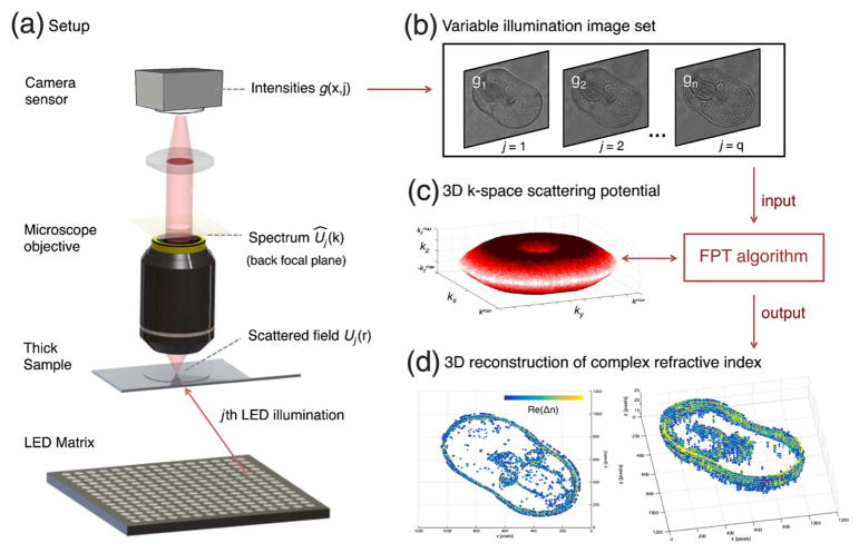
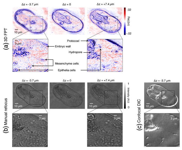
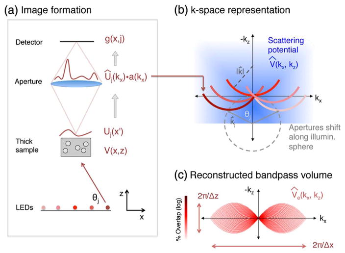
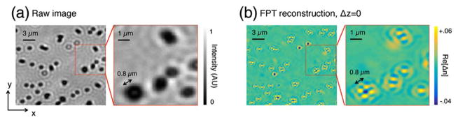
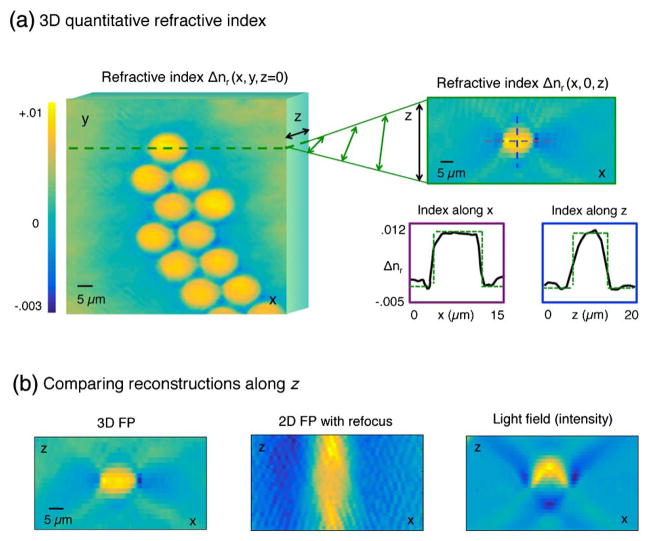
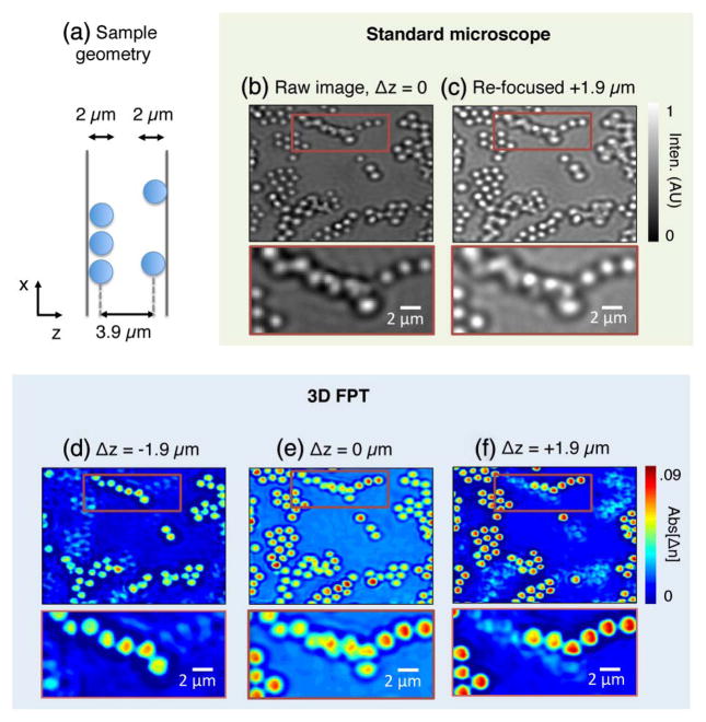
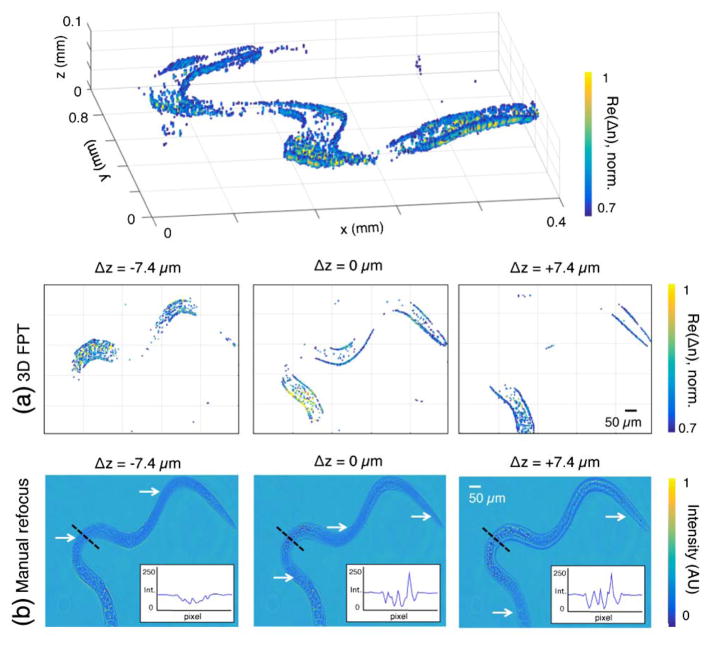
This paper presents a technique to image the complex index of refraction of a sample across three dimensions. The only required hardware is a standard microscope and an array of LEDs. The method, termed Fourier ptychographic tomography (FPT), first captures a sequence of intensity-only images of a sample under angularly varying illumination. Then, using principles from ptychography and diffraction tomography, it computationally solves for the sample structure in three dimensions. The experimental microscope demonstrates a lateral spatial resolution of 0.39 μm and an axial resolution of 3.7 μm at the Nyquist-Shannon sampling limit (0.54 and 5.0 μm at the Sparrow limit, respectively) across a total imaging depth of 110 μm. Unlike competing methods, this technique quantitatively measures the volumetric refractive index of primarily transparent and contiguous sample features without the need for interferometry or any moving parts. Wide field-of-view reconstructions of thick biological specimens suggest potential applications in pathology and developmental biology.

摘要

本文介绍了一种在三维空间对样品复折射率进行成像的技术。唯一所需的硬件是一台标准显微镜和一组发光二极管。该方法称为傅里叶叠层断层成像(FPT),首先在角度变化的照明条件下捕获样品的一系列仅强度图像。然后,利用叠层成像和衍射断层成像的原理,通过计算求解样品的三维结构。实验显微镜在110μm的总成像深度上,在奈奎斯特 - 香农采样极限下横向空间分辨率为0.39μm,轴向分辨率为3.7μm(在斯帕罗极限下分别为0.54μm和5.0μm)。与其他竞争方法不同,该技术无需干涉测量或任何移动部件,即可对主要为透明且连续的样品特征的体积折射率进行定量测量。厚生物标本的宽视场重建表明其在病理学和发育生物学方面具有潜在应用。

https://cdn.ncbi.nlm.nih.gov/pmc/blobs/0d56/5521281/ad5a2f484138/nihms835228f7.jpg
https://cdn.ncbi.nlm.nih.gov/pmc/blobs/0d56/5521281/3e326b8cbfcb/nihms835228f1.jpg
https://cdn.ncbi.nlm.nih.gov/pmc/blobs/0d56/5521281/896bf317a8fd/nihms835228f2.jpg
https://cdn.ncbi.nlm.nih.gov/pmc/blobs/0d56/5521281/f157263f5056/nihms835228f3.jpg
https://cdn.ncbi.nlm.nih.gov/pmc/blobs/0d56/5521281/b04bb79e5693/nihms835228f4.jpg
https://cdn.ncbi.nlm.nih.gov/pmc/blobs/0d56/5521281/46f39d5cc221/nihms835228f5.jpg
https://cdn.ncbi.nlm.nih.gov/pmc/blobs/0d56/5521281/13070717e550/nihms835228f6.jpg
https://cdn.ncbi.nlm.nih.gov/pmc/blobs/0d56/5521281/ad5a2f484138/nihms835228f7.jpg

相似文献

1
Diffraction tomography with Fourier ptychography.傅里叶叠层衍射层析成像
Optica. 2016 Aug;3(8):827-835. doi: 10.1364/OPTICA.3.000827. Epub 2016 Jul 27.
2
Multi-slice ptychographic tomography.多切片叠层相衬断层成像技术。
Sci Rep. 2018 Feb 1;8(1):2049. doi: 10.1038/s41598-018-20530-x.
3
Lens-free on-chip 3D microscopy based on wavelength-scanning Fourier ptychographic diffraction tomography.基于波长扫描傅里叶叠层衍射层析成像的无透镜片上三维显微镜技术。
Light Sci Appl. 2024 Sep 5;13(1):237. doi: 10.1038/s41377-024-01568-1.
4
Quantitative Jones matrix imaging using vectorial Fourier ptychography.使用矢量傅里叶叠层成像的定量琼斯矩阵成像
Biomed Opt Express. 2022 Feb 14;13(3):1457-1470. doi: 10.1364/BOE.448804. eCollection 2022 Mar 1.
5
Multiplexed coded illumination for Fourier Ptychography with an LED array microscope.用于基于LED阵列显微镜的傅里叶叠层成像术的多路复用编码照明
Biomed Opt Express. 2014 Jun 19;5(7):2376-89. doi: 10.1364/BOE.5.002376. eCollection 2014 Jul 1.
6
Content adaptive illumination for Fourier ptychography.用于傅里叶叠层成像的内容自适应照明
Opt Lett. 2014 Dec 1;39(23):6648-51. doi: 10.1364/OL.39.006648.
7
High-resolution fluorescence imaging via pattern-illuminated Fourier ptychography.通过图案照明傅里叶叠层成像实现高分辨率荧光成像。
Opt Express. 2014 Aug 25;22(17):20856-70. doi: 10.1364/OE.22.020856.
8
3D-coherent anti-Stokes Raman scattering Fourier ptychography tomography (CARS-FPT).三维相干反斯托克斯拉曼散射傅里叶叠层断层扫描(CARS-FPT)
Opt Express. 2021 Feb 1;29(3):4230-4239. doi: 10.1364/OE.416290.
9
Ptychographic microscope for three-dimensional imaging.用于三维成像的叠层成像显微镜。
Opt Express. 2014 May 19;22(10):12513-23. doi: 10.1364/OE.22.012513.
10
Near-field ptychographic microscope for quantitative phase imaging.
Opt Express. 2018 Sep 17;26(19):25471-25480. doi: 10.1364/OE.26.025471.

引用本文的文献

1
Inverse-scattering in biological samples via beam-propagation.通过光束传播对生物样本进行逆散射。
bioRxiv. 2025 Aug 22:2025.08.17.670744. doi: 10.1101/2025.08.17.670744.
2
Unified -space theory of optical coherence tomography.光学相干断层扫描的统一空间理论
Adv Opt Photonics. 2021 Jun 30;13(2):462-514. doi: 10.1364/aop.417102. Epub 2021 Jun 25.
3
From superior contrast to super resolution label free optical microscopy.从高对比度到超分辨率无标记光学显微镜。

本文引用的文献

1
Experimental robustness of Fourier ptychography phase retrieval algorithms.傅里叶叠层成像相位恢复算法的实验稳健性
Opt Express. 2015 Dec 28;23(26):33214-40. doi: 10.1364/OE.23.033214.
2
Solving ptychography with a convex relaxation.通过凸松弛解决叠层成像问题。
New J Phys. 2015 May;17. doi: 10.1088/1367-2630/17/5/053044.
3
Separation of three-dimensional scattering effects in tilt-series Fourier ptychography.倾斜序列傅里叶叠层成像中三维散射效应的分离
Npj Imaging. 2025 Jan 13;3(1):1. doi: 10.1038/s44303-024-00064-w.
4
Making sense of blobs, whorls, and shades: methods for label-free, inverse imaging in bright-field optical microscopy.理解斑点、漩涡和阴影:明场光学显微镜中无标记逆成像的方法。
Biophys Rev. 2025 Mar 18;17(2):335-345. doi: 10.1007/s12551-025-01301-1. eCollection 2025 Apr.
5
High-Fidelity Computational Microscopy via Feature-Domain Phase Retrieval.通过特征域相位恢复实现的高保真计算显微镜技术。
Adv Sci (Weinh). 2025 Jun;12(21):e2413975. doi: 10.1002/advs.202413975. Epub 2025 Feb 22.
6
Multi-modal transport of intensity diffraction tomography microscopy with an electrically tunable lens [Invited].基于电可调透镜的强度衍射层析显微镜多模态传输[特邀报告]
Biomed Opt Express. 2025 Jan 31;16(2):837-848. doi: 10.1364/BOE.545258. eCollection 2025 Feb 1.
7
Computational microscopy with coherent diffractive imaging and ptychography.基于相干衍射成像和叠层成像术的计算显微镜技术。
Nature. 2025 Jan;637(8045):281-295. doi: 10.1038/s41586-024-08278-z. Epub 2025 Jan 8.
8
Deciphering structural complexity of brain, joint, and muscle tissues using Fourier Ptychographic Scattered Light Microscopy.利用傅里叶叠层散斑显微镜解析脑、关节和肌肉组织的结构复杂性。
bioRxiv. 2025 Apr 9:2024.11.28.625428. doi: 10.1101/2024.11.28.625428.
9
Quadrant darkfield for label-free imaging of intracellular puncta.用于细胞内斑点无标记成像的象限暗场
J Biomed Opt. 2024 Nov;29(11):116501. doi: 10.1117/1.JBO.29.11.116501. Epub 2024 Nov 29.
10
Spatially-coded Fourier ptychography: flexible and detachable coded thin films for quantitative phase imaging with uniform phase transfer characteristics.空间编码傅里叶叠层成像术:用于具有均匀相位传递特性的定量相位成像的灵活且可拆卸编码薄膜
Adv Opt Mater. 2024 May 28;12(15). doi: 10.1002/adom.202303028. Epub 2024 Feb 7.
Ultramicroscopy. 2015 Nov;158:1-7. doi: 10.1016/j.ultramic.2015.06.010. Epub 2015 Jun 14.
4
Fourier ptychographic reconstruction using Wirtinger flow optimization.使用维特林格流优化的傅里叶叠层成像重建。
Opt Express. 2015 Feb 23;23(4):4856-66. doi: 10.1364/OE.23.004856.
5
High numerical aperture Fourier ptychography: principle, implementation and characterization.高数值孔径傅里叶叠层成像术:原理、实现与表征
Opt Express. 2015 Feb 9;23(3):3472-91. doi: 10.1364/OE.23.003472.
6
Optical imaging. Expansion microscopy.光学成像。扩展显微镜技术。
Science. 2015 Jan 30;347(6221):543-8. doi: 10.1126/science.1260088. Epub 2015 Jan 15.
7
Diffraction optical tomography using a quantitative phase imaging unit.使用定量相位成像单元的衍射光学层析成像。
Opt Lett. 2014 Dec 15;39(24):6935-8. doi: 10.1364/OL.39.006935.
8
Wide-field, high-resolution Fourier ptychographic microscopy.宽视场、高分辨率傅里叶叠层显微镜术
Nat Photonics. 2013 Sep 1;7(9):739-745. doi: 10.1038/nphoton.2013.187.
9
Multiplexed coded illumination for Fourier Ptychography with an LED array microscope.用于基于LED阵列显微镜的傅里叶叠层成像术的多路复用编码照明
Biomed Opt Express. 2014 Jun 19;5(7):2376-89. doi: 10.1364/BOE.5.002376. eCollection 2014 Jul 1.
10
Ptychographic microscope for three-dimensional imaging.用于三维成像的叠层成像显微镜。
Opt Express. 2014 May 19;22(10):12513-23. doi: 10.1364/OE.22.012513.