Thivolet Charles, Vial Guillaume, Cassel Romeo, Rieusset Jennifer, Madec Anne-Marie
INSERM UMR-1060, CarMeN Laboratory, Lyon 1 University, INRA U1235, Lyon, France.
Hospices Civils de Lyon, Lyon-Sud Hospital, Department of Endocrinology and Diabetes, Pierre-Bénite, France.
PLoS One. 2017 Jul 25;12(7):e0182027. doi: 10.1371/journal.pone.0182027. eCollection 2017.
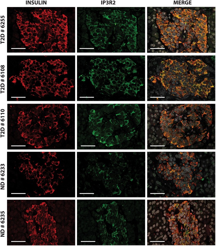
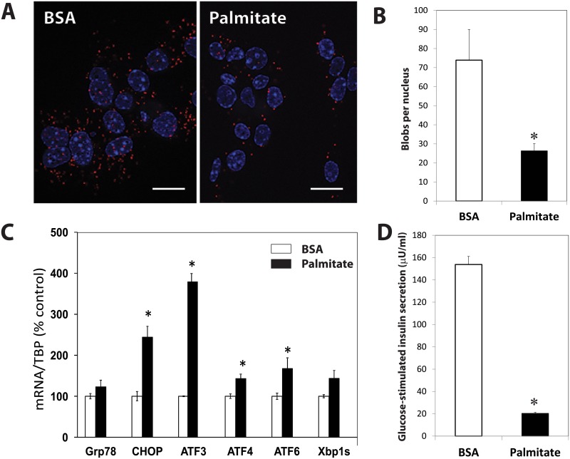
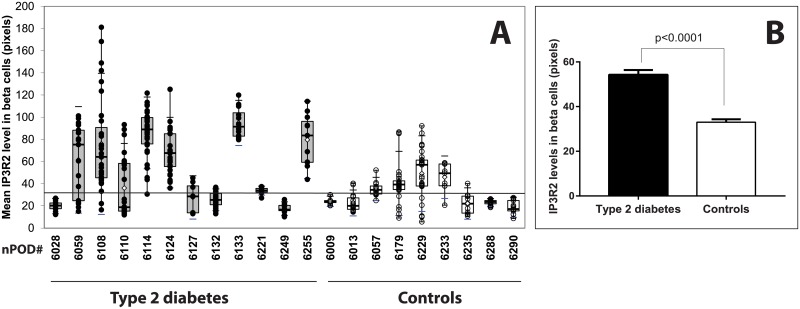
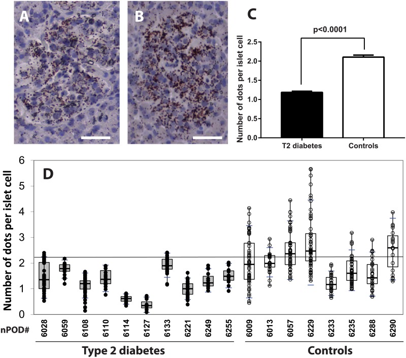
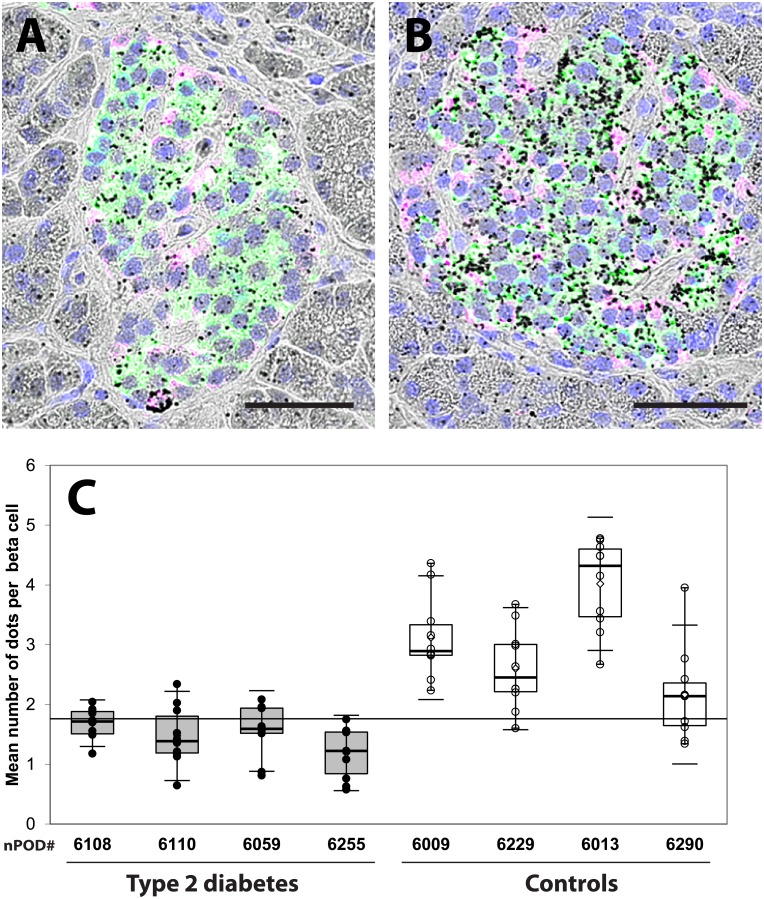
Type 2 diabetes develops when beta cells are not able to fulfill insulin needs. The role of the endoplasmic reticulum-mitochondria junction in coordinating the functions of these two organelles throughout the natural history of type 2 diabetes is determinant and may explain the alterations of insulin biosynthesis. Our goal was to study endoplasmic reticulum and mitochondrial interactions in human beta cells from organ donors with type 2 diabetes. Pancreas samples were obtained via the network for pancreatic organ donors with diabetes (nPOD) based on disease status with 12 subjects with type 2 diabetes and 9 non-diabetic controls. We examined pancreatic specimens by immunofluorescence, in situ hybridization and in situ proximity ligation assay and compared the results to an in vitro model of beta-cell dysfunction. Expression of proteins that enable tethering and exchanges between endoplasmic reticulum (ER) and mitochondria and quantification of interconnection through mitochondria associated membranes (MAM) was investigated. In beta cells from type 2 diabetic cases as compared to controls, there was a significant increase in reticular expression of inositol triphosphate receptor-2 (IP3R2) both at the protein and mRNA levels, no difference in mitochondrial transit peptide receptor TOM20 and mitofusin-2 expressions, and a decrease in the expression of voltage-dependent anion channel-1 (VDAC-1). The number of IP3R2-VDAC-1 complexes identified by in situ proximity ligation assay was significantly lower in diabetic islets and in beta cells of diabetics as compared to controls. Treatment of Min6-B1 cells with palmitate altered glucose-stimulated insulin secretion, increased ER stress and significantly reduced ER-mitochondrial interactions. We can conclude that specific changes in reticular and mitochondrial beta cell proteins characterize human type 2 diabetes with reduction in organelle interactions. This finding opens new targets of intervention.
当β细胞无法满足胰岛素需求时,2型糖尿病就会发生。内质网 - 线粒体连接在2型糖尿病的整个自然病程中协调这两个细胞器功能的作用是决定性的,并且可能解释胰岛素生物合成的改变。我们的目标是研究2型糖尿病器官供体的人β细胞中的内质网和线粒体相互作用。通过糖尿病胰腺器官供体网络(nPOD)根据疾病状态获取胰腺样本,其中有12名2型糖尿病患者和9名非糖尿病对照。我们通过免疫荧光、原位杂交和原位邻近连接分析检查胰腺标本,并将结果与β细胞功能障碍的体外模型进行比较。研究了在内质网(ER)和线粒体之间实现拴系和交换的蛋白质表达以及通过线粒体相关膜(MAM)的互连定量。与对照组相比,2型糖尿病患者的β细胞中,肌醇三磷酸受体2(IP3R2)在蛋白质和mRNA水平上的网状表达均显著增加,线粒体转运肽受体TOM20和线粒体融合蛋白2的表达没有差异,而电压依赖性阴离子通道1(VDAC - 1)的表达降低。与对照组相比,通过原位邻近连接分析鉴定的IP3R2 - VDAC - 1复合物的数量在糖尿病胰岛和糖尿病患者的β细胞中显著降低。用棕榈酸处理Min6 - B1细胞会改变葡萄糖刺激的胰岛素分泌,增加内质网应激并显著减少内质网 - 线粒体相互作用。我们可以得出结论,网状和线粒体β细胞蛋白的特定变化是人类2型糖尿病的特征,细胞器相互作用减少。这一发现开辟了新的干预靶点。