Jana Manisha, Nair Nikhil, Gupta Arun K, Kabra Madhulika, Gupta Neerja
Department of Radiodiagnosis, All India Institute of Medical Sciences, New Delhi, India.
Department of Pediatrics, All India Institute of Medical Sciences, New Delhi, India.
Indian J Radiol Imaging. 2017 Apr-Jun;27(2):187-199. doi: 10.4103/ijri.IJRI_367_16.
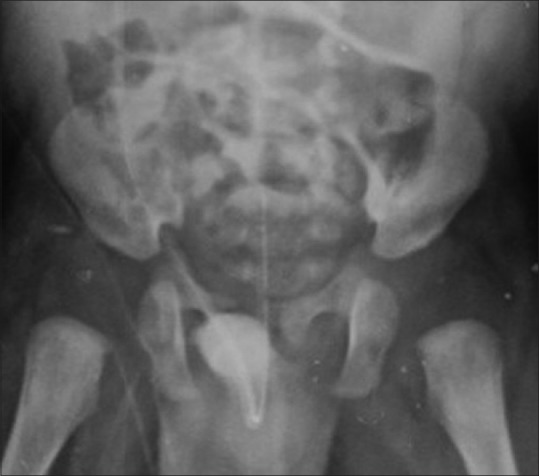
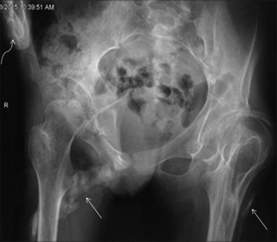
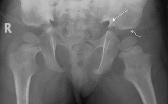
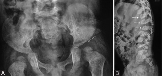
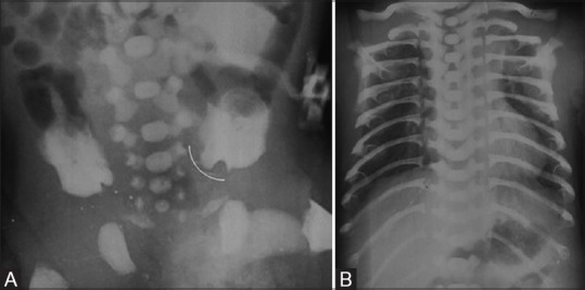
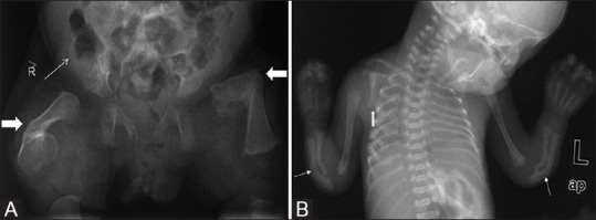
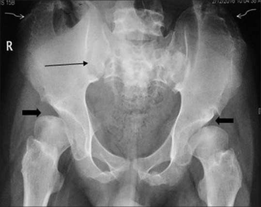
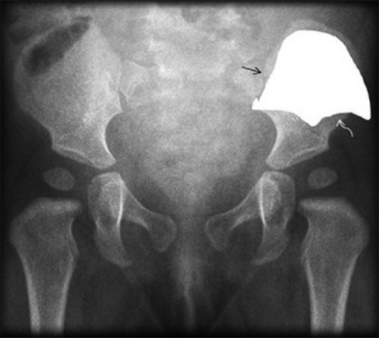
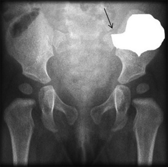
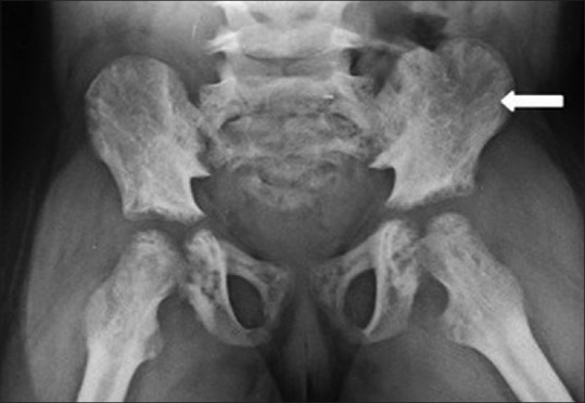
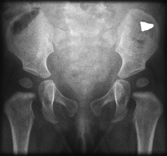
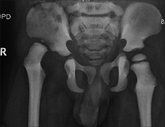
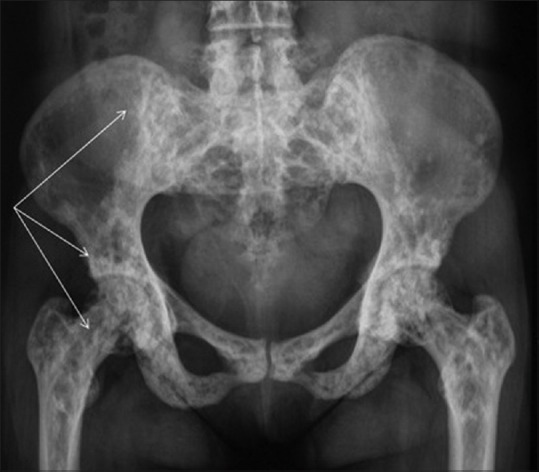
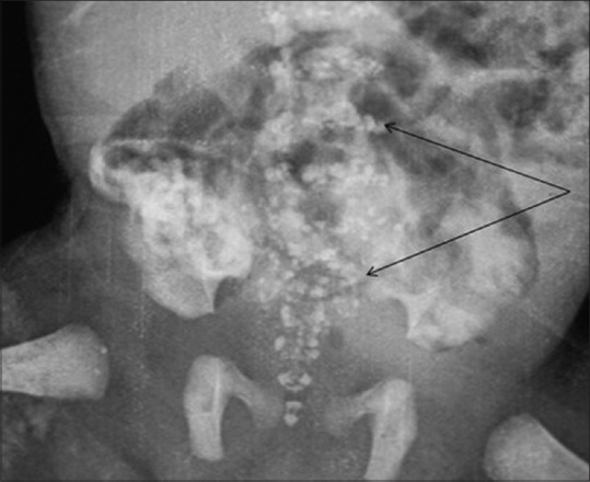
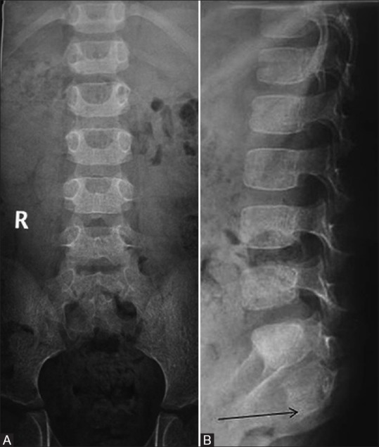
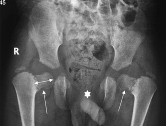
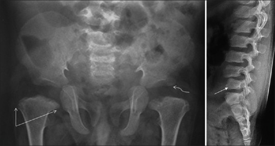
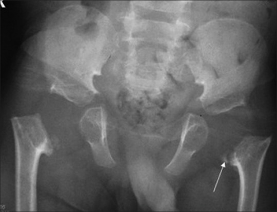
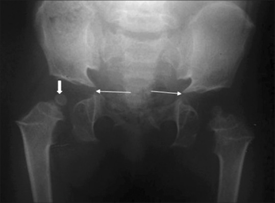
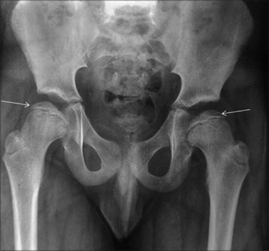
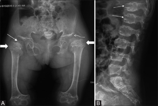
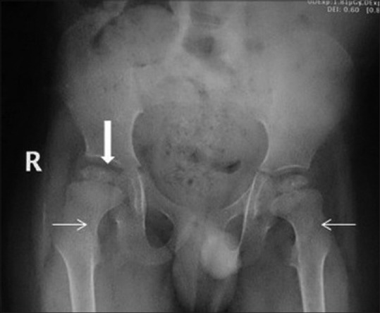
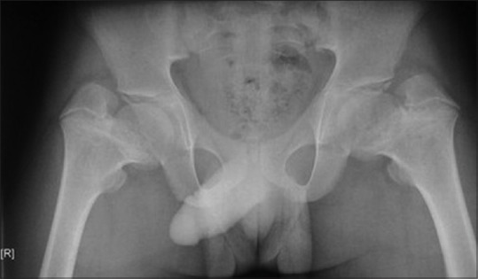
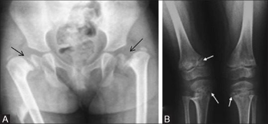
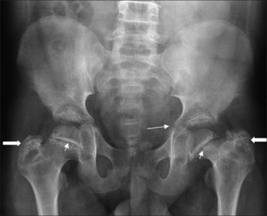
The bony pelvis is constituted by the ilium, ischium, pubis, and sacrum. The pelvic radiograph is an important component of the skeletal survey performed in suspected skeletal dysplasia. Most of the common skeletal dysplasias have either minor or major radiological abnormalities; hence, knowledge of the normal radiological appearance of bony pelvis is vital for recognizing the early signs of various skeletal dysplasias. This article discusses many common and some uncommon radiological findings on pelvic radiographs along with the specific dysplasia in which they are seen; common differential diagnostic considerations are also discussed.
骨盆由髂骨、坐骨、耻骨和骶骨组成。骨盆X线片是疑似骨骼发育异常患者进行骨骼检查的重要组成部分。大多数常见的骨骼发育异常都有轻微或严重的放射学异常;因此,了解骨盆的正常放射学表现对于识别各种骨骼发育异常的早期迹象至关重要。本文讨论了骨盆X线片上许多常见和一些不常见的放射学表现及其所对应的特定发育异常;还讨论了常见的鉴别诊断要点。