• 文献检索
  • 文档翻译
  • 深度研究
  • 学术资讯
  • Suppr Zotero 插件Zotero 插件
  • 邀请有礼
  • 套餐&价格
  • 历史记录
应用&插件
Suppr Zotero 插件Zotero 插件浏览器插件Mac 客户端Windows 客户端微信小程序
定价
高级版会员购买积分包购买API积分包
服务
文献检索文档翻译深度研究API 文档MCP 服务
关于我们
关于 Suppr公司介绍联系我们用户协议隐私条款
关注我们

Suppr 超能文献

核心技术专利:CN118964589B侵权必究
粤ICP备2023148730 号-1Suppr @ 2026

文献检索

告别复杂PubMed语法,用中文像聊天一样搜索,搜遍4000万医学文献。AI智能推荐,让科研检索更轻松。

立即免费搜索

文件翻译

保留排版,准确专业,支持PDF/Word/PPT等文件格式,支持 12+语言互译。

免费翻译文档

深度研究

AI帮你快速写综述,25分钟生成高质量综述,智能提取关键信息,辅助科研写作。

立即免费体验

指数表观扩散系数在3.0特斯拉扩散加权磁共振成像对乳腺病变特征性分析中的作用

Role of exponential apparent diffusion coefficient in characterizing breast lesions by 3.0 Tesla diffusion-weighted magnetic resonance imaging.

作者信息

Kothari Shweta, Singh Archana, Das Utpalendu, Sarkar Diptendra K, Datta Chhanda, Hazra Avijit

机构信息

Department of Radio Diagnosis, IPGME and R and SSKM Hospital, Kolkata, West Bengal, India.

Department of Surgery, IPGME and R and SSKM Hospital, Kolkata, West Bengal, India.

出版信息

Indian J Radiol Imaging. 2017 Apr-Jun;27(2):229-236. doi: 10.4103/ijri.IJRI_405_16.

DOI:10.4103/ijri.IJRI_405_16
PMID:28744085
原文链接:https://pmc.ncbi.nlm.nih.gov/articles/PMC5510322/
Abstract

OBJECTIVE

To evaluate the role of exponential apparent diffusion coefficient (ADC) as a tool for differentiating benign and malignant breast lesions.

PATIENTS AND METHODS

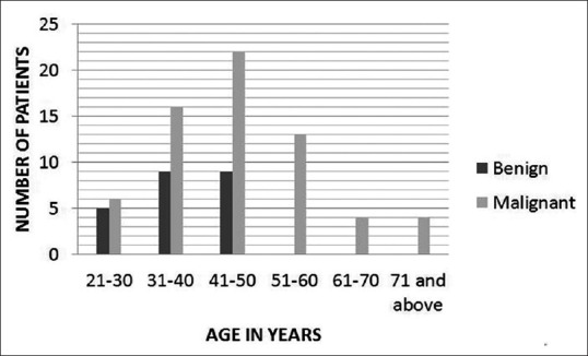
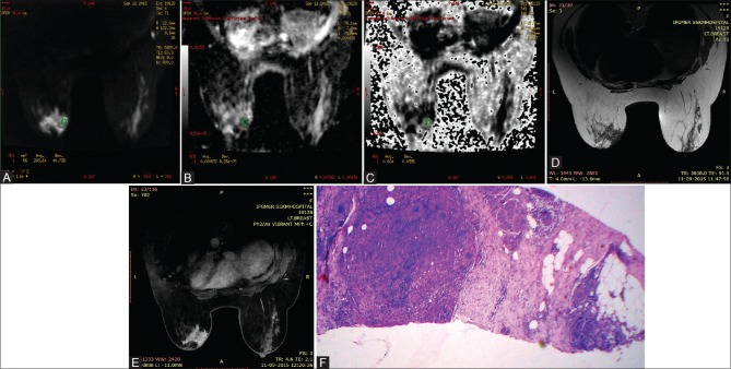
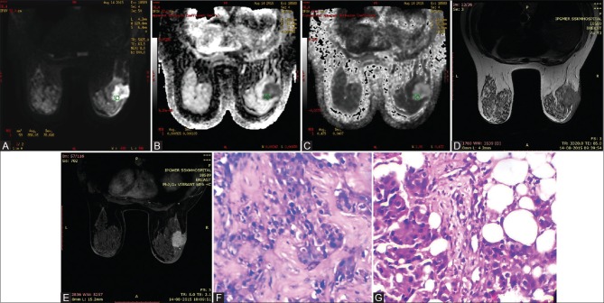
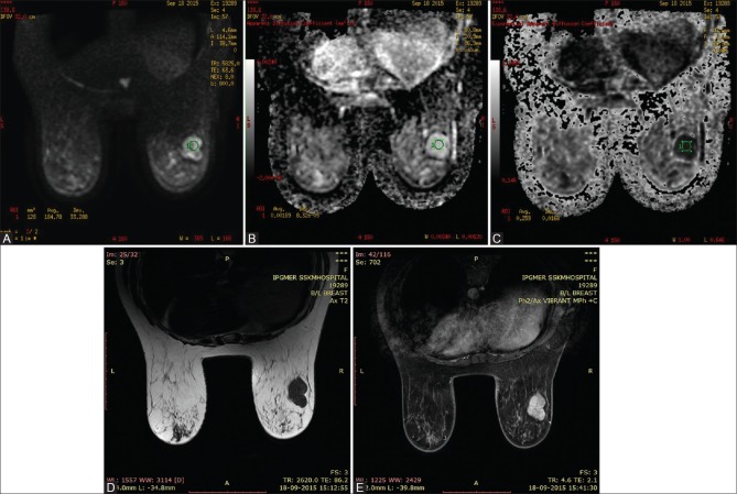
This prospective observational study included 88 breast lesions in 77 patients (between 18 and 85 years of age) who underwent 3T breast magnetic resonance imaging (MRI) including diffusion-weighted imaging (DWI) using b-values of 0 and 800 s/mm before biopsy. Mean exponential ADC and ADC of benign and malignant lesions obtained from DWI were compared. Receiver operating characteristics (ROC) curve analysis was undertaken to identify any cut-off for exponential ADC and ADC to predict malignancy. value of <0.05 was considered statistically significant. Histopathology was taken as the gold standard.

RESULTS

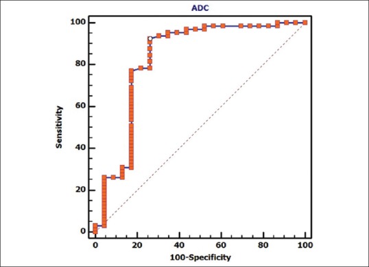
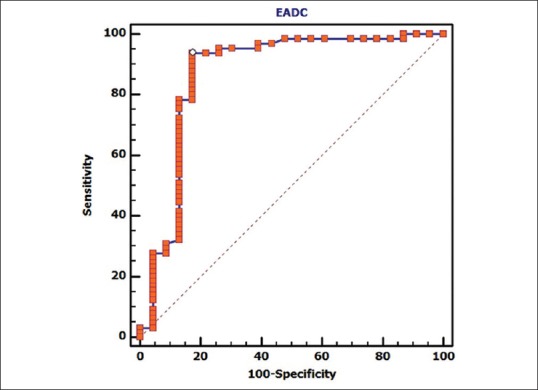
According to histopathology, 65 lesions were malignant and 23 were benign. The mean ADC and exponential ADC values of malignant lesions were 0.9526 ± 0.203 × 10 mm/s and 0.4774 ± 0.071, respectively, and for benign lesions were 1.48 ± 0.4903 × 10 mm/s and 0.317 ± 0.1152, respectively. For both the parameters, differences were highly significant ( < 0.001). Cut-off value of ≤0.0011 mm/s ( < 0.0001) for ADC provided 92.3% sensitivity and 73.9% specificity, whereas with an exponential ADC cut-off value of >0.4 ( < 0.0001) for malignant lesions, 93.9% sensitivity and 82.6% specificity was obtained. The performance of ADC and exponential ADC in distinguishing benign and malignant breast lesions based on respective cut-offs was comparable ( = 0.109).

CONCLUSION

Exponential ADC can be used as a quantitative adjunct tool for characterizing breast lesions with comparable sensitivity and specificity as that of ADC.

摘要

目的

评估指数表观扩散系数(ADC)作为鉴别乳腺良恶性病变工具的作用。

患者与方法

这项前瞻性观察性研究纳入了77例患者(年龄在18至85岁之间)的88个乳腺病变,这些患者在活检前接受了3T乳腺磁共振成像(MRI),包括使用b值为0和800 s/mm²的扩散加权成像(DWI)。比较了从DWI获得的良性和恶性病变的平均指数ADC和ADC。进行了受试者操作特征(ROC)曲线分析,以确定指数ADC和ADC预测恶性肿瘤的任何临界值。P值<0.05被认为具有统计学意义。组织病理学被视为金标准。

结果

根据组织病理学,65个病变为恶性,23个为良性。恶性病变的平均ADC值和指数ADC值分别为0.9526±0.203×10⁻³mm²/s和0.4774±0.071,良性病变分别为1.48±0.4903×10⁻³mm²/s和0.317±0.1152。对于这两个参数,差异均具有高度统计学意义(P<0.001)。ADC的临界值≤0.0011×10⁻³mm²/s(P<0.0001)时,敏感性为92.3%,特异性为73.9%;而对于恶性病变,指数ADC临界值>0.4(P<0.0001)时,敏感性为93.9%,特异性为82.6%。基于各自临界值,ADC和指数ADC在区分乳腺良恶性病变方面的表现相当(P = 0.109)。

结论

指数ADC可作为一种定量辅助工具,用于表征乳腺病变,其敏感性和特异性与ADC相当。

https://cdn.ncbi.nlm.nih.gov/pmc/blobs/083b/5510322/e2e0fab56796/IJRI-27-229-g009.jpg
https://cdn.ncbi.nlm.nih.gov/pmc/blobs/083b/5510322/ba9df709b007/IJRI-27-229-g001.jpg
https://cdn.ncbi.nlm.nih.gov/pmc/blobs/083b/5510322/db33dfc2e776/IJRI-27-229-g002.jpg
https://cdn.ncbi.nlm.nih.gov/pmc/blobs/083b/5510322/2f11e562122b/IJRI-27-229-g003.jpg
https://cdn.ncbi.nlm.nih.gov/pmc/blobs/083b/5510322/ad1bd11905b0/IJRI-27-229-g004.jpg
https://cdn.ncbi.nlm.nih.gov/pmc/blobs/083b/5510322/18d54e2d5d5a/IJRI-27-229-g005.jpg
https://cdn.ncbi.nlm.nih.gov/pmc/blobs/083b/5510322/a71dddb85d99/IJRI-27-229-g008.jpg
https://cdn.ncbi.nlm.nih.gov/pmc/blobs/083b/5510322/e2e0fab56796/IJRI-27-229-g009.jpg

相似文献

1
Role of exponential apparent diffusion coefficient in characterizing breast lesions by 3.0 Tesla diffusion-weighted magnetic resonance imaging.指数表观扩散系数在3.0特斯拉扩散加权磁共振成像对乳腺病变特征性分析中的作用
Indian J Radiol Imaging. 2017 Apr-Jun;27(2):229-236. doi: 10.4103/ijri.IJRI_405_16.
2
Comparison of the diagnostic performances of diffusion parameters in diffusion weighted imaging and diffusion tensor imaging of breast lesions.扩散加权成像和扩散张量成像中扩散参数对乳腺病变诊断性能的比较。
Eur J Radiol. 2013 Dec;82(12):e801-6. doi: 10.1016/j.ejrad.2013.09.001. Epub 2013 Sep 13.
3
Diffusion kurtosis imaging does not improve differentiation performance of breast lesions in a short clinical protocol.扩散峰度成像在短期临床方案中不能提高乳腺病变的鉴别性能。
Magn Reson Imaging. 2019 Nov;63:205-216. doi: 10.1016/j.mri.2019.08.007. Epub 2019 Aug 16.
4
Breast lesion characterization using whole-lesion histogram analysis with stretched-exponential diffusion model.使用拉伸指数扩散模型的全病变直方图分析进行乳腺病变特征描述。
J Magn Reson Imaging. 2018 Jun;47(6):1701-1710. doi: 10.1002/jmri.25904. Epub 2017 Nov 22.
5
Diagnostic value of diffusion-weighted magnetic resonance imaging: differentiation of benign and malignant lymph nodes in different regions of the body.磁共振扩散加权成像的诊断价值:身体不同区域良恶性淋巴结的鉴别
Clin Imaging. 2015 Sep-Oct;39(5):856-62. doi: 10.1016/j.clinimag.2015.05.006. Epub 2015 May 22.
6
Diffusion weighted imaging and apparent diffusion coefficient in 3 tesla magnetic resonance imaging of breast lesions.3特斯拉磁共振成像中乳腺病变的扩散加权成像及表观扩散系数
Cancer Invest. 2015 May;33(5):159-64. doi: 10.3109/07357907.2015.1019674. Epub 2015 Apr 1.
7
Diffusion-weighted imaging: determination of the best pair of b-values to discriminate breast lesions.扩散加权成像:确定用于鉴别乳腺病变的最佳b值对。
Br J Radiol. 2014 Jul;87(1039):20130807. doi: 10.1259/bjr.20130807. Epub 2014 May 16.
8
Added Value of Diffusion-Weighted Magnetic Resonance Imaging in Differentiating Musculoskeletal Tumors Using Sensitivity and Specificity: A Retrospective Study and Review of Literature.利用敏感性和特异性评估扩散加权磁共振成像在鉴别肌肉骨骼肿瘤中的附加价值:一项回顾性研究及文献综述
Cureus. 2021 Jan 1;13(1):e12422. doi: 10.7759/cureus.12422.
9
Differentiation of Breast Lesions and Distinguishing Their Histological Subtypes Using Diffusion-Weighted Imaging and ADC Values.利用扩散加权成像和表观扩散系数值鉴别乳腺病变及其组织学亚型
Front Oncol. 2020 Mar 13;10:332. doi: 10.3389/fonc.2020.00332. eCollection 2020.
10
Application Value of Mathematical Models of Diffusion-Weighted Magnetic Resonance Imaging in Differentiating Breast Cancer Lesions.扩散加权磁共振成像数学模型在鉴别乳腺癌病变中的应用价值
Evid Based Complement Alternat Med. 2021 Aug 28;2021:1481271. doi: 10.1155/2021/1481271. eCollection 2021.

引用本文的文献

1
A Prospective Observational Study of Diagnostic Reliability of Semiquantitative and Quantitative High b-Value Diffusion-Weighted MRI in Distinguishing between Benign and Malignant Lung Lesions at 3 Tesla.一项关于在3特斯拉场强下,半定量和定量高b值扩散加权磁共振成像鉴别肺良恶性病变诊断可靠性的前瞻性观察研究。
Indian J Radiol Imaging. 2023 Aug 16;34(1):6-15. doi: 10.1055/s-0043-1771530. eCollection 2024 Jan.
2
Diagnostic performance of breast tumor tissue selection in diffusion weighted imaging: A systematic review and meta-analysis.扩散加权成像中乳腺肿瘤组织选择的诊断性能:系统评价和荟萃分析。
PLoS One. 2020 May 6;15(5):e0232856. doi: 10.1371/journal.pone.0232856. eCollection 2020.

本文引用的文献

1
Qualitative and quantitative diffusion-weighted imaging of the breast at 3T - A useful adjunct to contrast-enhanced MRI in characterization of breast lesions.3T 乳腺的定性和定量扩散加权成像——对比增强 MRI 在乳腺病变特征描述中的有用辅助手段
Indian J Radiol Imaging. 2015 Oct-Dec;25(4):397-403. doi: 10.4103/0971-3026.169455.
2
Exponential apparent diffusion coefficient in evaluating prostate cancer at 3 T: preliminary experience.3T场强下指数表观扩散系数在前列腺癌评估中的初步经验
Br J Radiol. 2016;89(1058):20150470. doi: 10.1259/bjr.20150470. Epub 2015 Dec 10.
3
Breast magnetic resonance imaging performance: safety, techniques, and updates on diffusion-weighted imaging and magnetic resonance spectroscopy.
乳腺磁共振成像性能:安全性、技术以及扩散加权成像和磁共振波谱的进展
Top Magn Reson Imaging. 2014 Dec;23(6):373-84. doi: 10.1097/RMR.0000000000000035.
4
Differentiation between benign and malignant breast lesions using quantitative diffusion-weighted sequence on 3 T MRI.使用 3T MRI 上的定量扩散加权序列对乳腺良恶性病变进行鉴别。
Clin Radiol. 2014 Jan;69(1):63-71. doi: 10.1016/j.crad.2013.08.007. Epub 2013 Oct 22.
5
EADC Values in Diagnosis of Renal Lesions by 3.0 T Diffusion-Weighted Magnetic Resonance Imaging: Compared with the ADC Values.3.0 T扩散加权磁共振成像中表观扩散系数值在肾病变诊断中的应用:与扩散系数值的比较
Appl Magn Reson. 2013 Mar;44(3):349-363. doi: 10.1007/s00723-012-0376-z. Epub 2012 Jul 5.
6
Understanding the mathematics involved in calculating apparent diffusion coefficient maps.理解计算表观扩散系数图所涉及的数学知识。
AJR Am J Roentgenol. 2012 Dec;199(6):W784. doi: 10.2214/AJR.12.9231.
7
Diffusion-weighted imaging of the breast: principles and clinical applications.乳腺扩散加权成像:原理与临床应用。
Radiographics. 2011 Jul-Aug;31(4):1059-84. doi: 10.1148/rg.314105160.
8
Contribution of diffusion-weighted imaging to dynamic contrast-enhanced MRI in the characterization of breast tumors.扩散加权成像对动态对比增强 MRI 在乳腺肿瘤特征描述中的作用。
AJR Am J Roentgenol. 2011 Jan;196(1):210-7. doi: 10.2214/AJR.10.4258.
9
Quantitative diffusion weighted imaging for differentiation of benign and malignant breast lesions: the influence of the choice of b-values.定量扩散加权成像鉴别乳腺良恶性病变:b 值选择的影响。
J Magn Reson Imaging. 2010 May;31(5):1100-5. doi: 10.1002/jmri.22152.
10
The role of parallel diffusion-weighted imaging and apparent diffusion coefficient (ADC) map values for evaluating breast lesions: preliminary results.平行弥散加权成像和表观扩散系数(ADC)图值在评价乳腺病变中的作用:初步结果。
Acad Radiol. 2010 Apr;17(4):456-63. doi: 10.1016/j.acra.2009.12.004.