Department of Radiology, University of Groningen, University Medical Center Groningen, Groningen, The Netherlands.
Department of Radiology, Utrecht University, University Medical Center Utrecht, Utrecht, The Netherlands.
PLoS One. 2020 May 6;15(5):e0232856. doi: 10.1371/journal.pone.0232856. eCollection 2020.
Several methods for tumor delineation are used in literature on breast diffusion weighted imaging (DWI) to measure the apparent diffusion coefficient (ADC). However, in the process of reaching consensus on breast DWI scanning protocol, image analysis and interpretation, still no standardized optimal breast tumor tissue selection (BTTS) method exists. Therefore, the purpose of this study is to assess the impact of BTTS methods on ADC in the discrimination of benign from malignant breast lesions in DWI in terms of sensitivity, specificity and area under the curve (AUC).
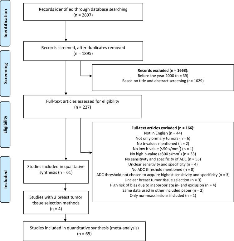
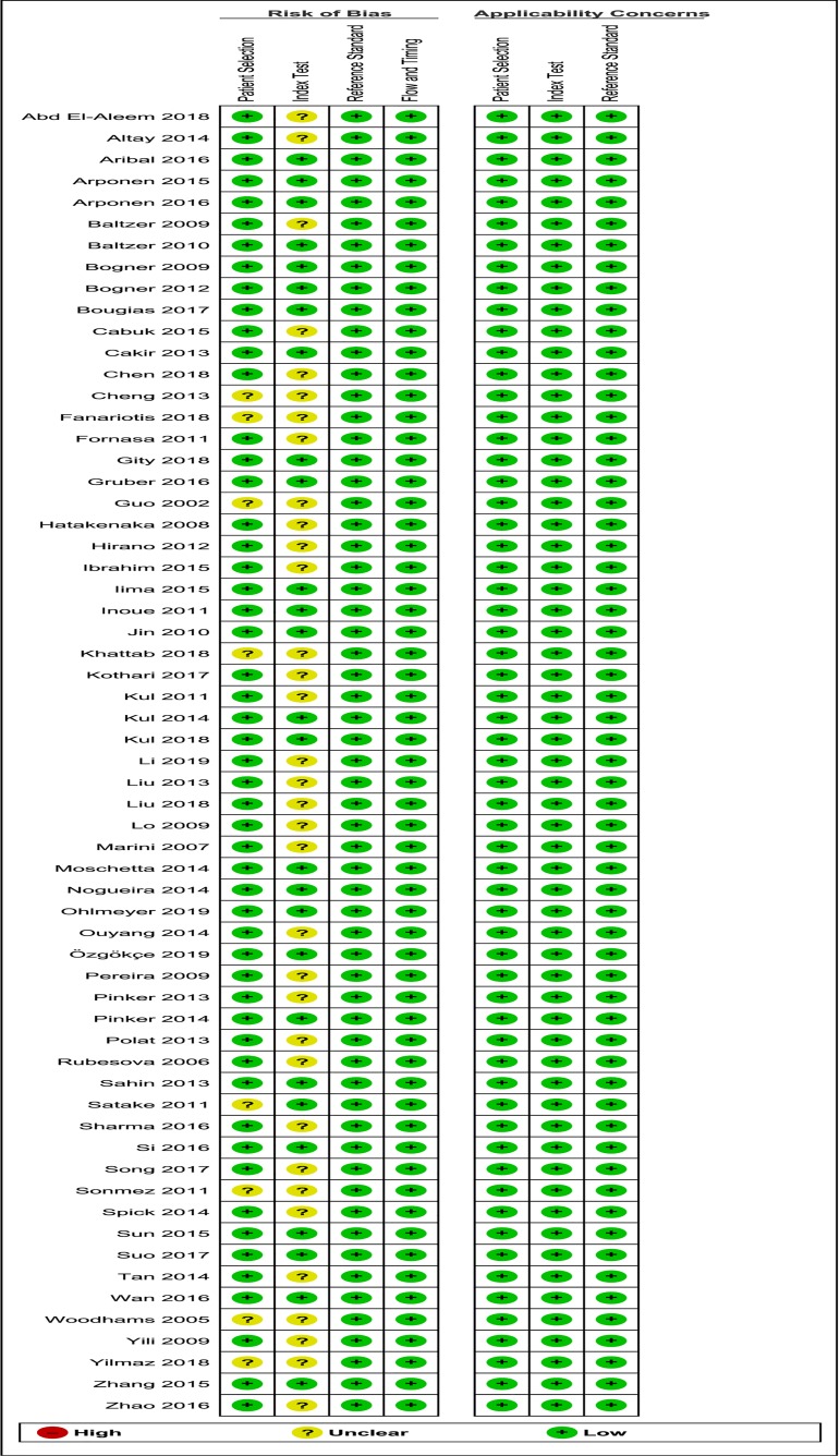
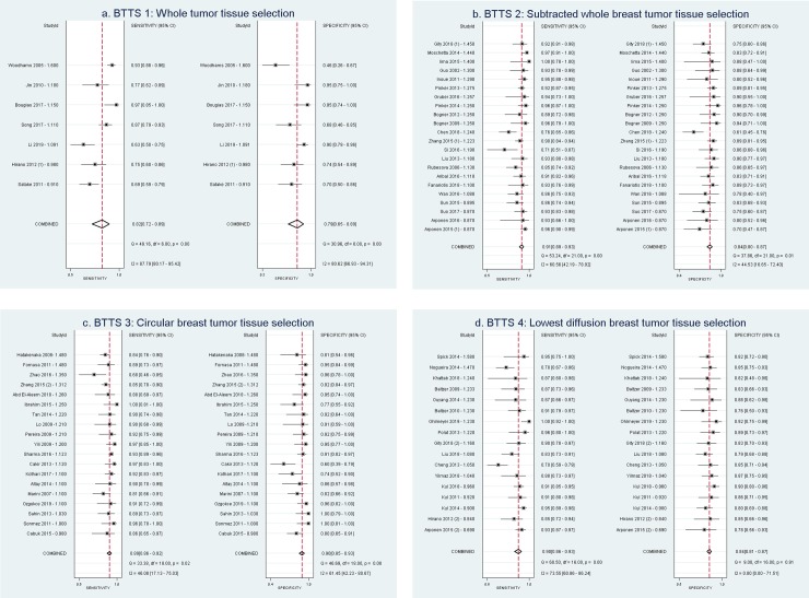
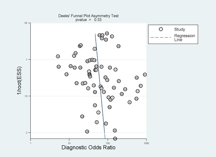
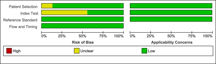
In this systematic review and meta-analysis, adhering to the PRISMA statement, 61 studies, with 65 study subsets, in females with benign or malignant primary breast lesions (6291 lesions) were assessed. Studies on DWI, quantified by ADC, scanned on 1.5 and 3.0 Tesla and using b-values 0/50 and ≥ 800 s/mm2 were included. PubMed and EMBASE were searched for studies up to 23-10-2019 (n = 2897). Data were pooled based on four BTTS methods (by definition of measured region of interest, ROI): BTTS1: whole breast tumor tissue selection, BTTS2: subtracted whole breast tumor tissue selection, BTTS3: circular breast tumor tissue selection and BTTS4: lowest diffusion breast tumor tissue selection. BTTS methods 2 and 3 excluded necrotic, cystic and hemorrhagic areas. Pooled sensitivity, specificity and AUC of the BTTS methods were calculated. Heterogeneity was explored using the inconsistency index (I2) and considering covariables: field strength, lowest b-value, image of BTTS selection, pre-or post-contrast DWI, slice thickness and ADC threshold. Pooled sensitivity, specificity and AUC were: 0.82 (0.72-0.89), 0.79 (0.65-0.89), 0.88 (0.85-0.90) for BTTS1; 0.91 (0.89-0.93), 0.84 (0.80-0.87), 0.94 (0.91-0.96) for BTTS2; 0.89 (0.86-0.92), 0.90 (0.85-0.93), 0.95 (0.93-0.96) for BTTS3 and 0.90 (0.86-0.93), 0.84 (0.81-0.87), 0.86 (0.82-0.88) for BTTS4, respectively. Significant heterogeneity was found between studies (I2 = 95).
None of the breast tissue selection (BTTS) methodologies outperformed in differentiating benign from malignant breast lesions. The high heterogeneity of ADC data acquisition demands further standardization, such as DWI acquisition parameters and tumor tissue selection to substantially increase the reliability of DWI of the breast.
在关于乳腺弥散加权成像(DWI)的文献中,有几种用于肿瘤勾画的方法来测量表观扩散系数(ADC)。然而,在就乳腺 DWI 扫描方案、图像分析和解释达成共识的过程中,仍然没有标准化的最佳乳腺肿瘤组织选择(BTTS)方法。因此,本研究的目的是评估 BTTS 方法对 DWI 中良恶性乳腺病变鉴别诊断的 ADC 的影响,从灵敏度、特异性和曲线下面积(AUC)方面进行评估。
在本系统评价和荟萃分析中,我们遵循 PRISMA 声明,评估了 61 项研究,其中包括 65 个研究亚组,研究对象为女性良性或恶性原发性乳腺病变(6291 个病变)。包括使用 0/50 和≥800 s/mm2 b 值进行 DWI 扫描的 1.5 和 3.0T 研究。我们在 PubMed 和 EMBASE 上搜索了截至 2019 年 10 月 23 日的研究(n=2897)。根据四种 BTTS 方法(通过测量感兴趣区域 ROI 的定义)进行数据汇总:BTTS1:全乳腺肿瘤组织选择;BTTS2:减去全乳腺肿瘤组织选择;BTTS3:圆形乳腺肿瘤组织选择;BTTS4:最低扩散乳腺肿瘤组织选择。BTTS 方法 2 和 3 排除了坏死、囊性和出血区域。计算了 BTTS 方法的汇总敏感性、特异性和 AUC。使用不一致指数(I2)并考虑协变量(场强、最低 b 值、BTTS 选择图像、对比前/后 DWI、切片厚度和 ADC 阈值)来探索异质性。汇总敏感性、特异性和 AUC 分别为:BTTS1:0.82(0.72-0.89)、0.79(0.65-0.89)、0.88(0.85-0.90);BTTS2:0.91(0.89-0.93)、0.84(0.80-0.87)、0.94(0.91-0.96);BTTS3:0.89(0.86-0.92)、0.90(0.85-0.93)、0.95(0.93-0.96);BTTS4:0.90(0.86-0.93)、0.84(0.81-0.87)、0.86(0.82-0.88)。研究之间存在显著的异质性(I2=95)。
在鉴别良恶性乳腺病变方面,没有一种乳腺组织选择(BTTS)方法具有优势。ADC 数据采集的高度异质性需要进一步标准化,例如 DWI 采集参数和肿瘤组织选择,以极大地提高乳腺 DWI 的可靠性。