• 文献检索
  • 文档翻译
  • 深度研究
  • 学术资讯
  • Suppr Zotero 插件Zotero 插件
  • 邀请有礼
  • 套餐&价格
  • 历史记录
应用&插件
Suppr Zotero 插件Zotero 插件浏览器插件Mac 客户端Windows 客户端微信小程序
定价
高级版会员购买积分包购买API积分包
服务
文献检索文档翻译深度研究API 文档MCP 服务
关于我们
关于 Suppr公司介绍联系我们用户协议隐私条款
关注我们

Suppr 超能文献

核心技术专利:CN118964589B侵权必究
粤ICP备2023148730 号-1Suppr @ 2026

文献检索

告别复杂PubMed语法,用中文像聊天一样搜索,搜遍4000万医学文献。AI智能推荐,让科研检索更轻松。

立即免费搜索

文件翻译

保留排版,准确专业,支持PDF/Word/PPT等文件格式,支持 12+语言互译。

免费翻译文档

深度研究

AI帮你快速写综述,25分钟生成高质量综述,智能提取关键信息,辅助科研写作。

立即免费体验

剪切波到达时间轮廓显示在剪切波弹性成像中的价值用于乳腺肿块的诊断。

Value of shear wave arrival time contour display in shear wave elastography for breast masses diagnosis.

机构信息

Department of Medical Ultrasound, Shanghai Tenth People's Hospital, Ultrasound Research and Education Institute, Tongji University School of Medicine, Shanghai, 200072, China.

Thyroid Institute, Tongji University School of Medicine, Shanghai, 200072, China.

出版信息

Sci Rep. 2017 Aug 1;7(1):7036. doi: 10.1038/s41598-017-07389-0.

DOI:10.1038/s41598-017-07389-0
PMID:28765627
原文链接:https://pmc.ncbi.nlm.nih.gov/articles/PMC5539095/
Abstract

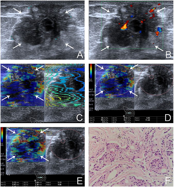
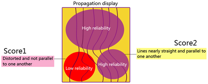
To evaluate the diagnostic performance of shear wave arrival time contour (SWATC) display for the diagnosis of breast lesions and to identify factors associated with the quality of shear wave propagation (QSWP) in breast lesions. This study included 277 pathologically confirmed breast lesions. Conventional B-mode ultrasound characteristics and shear wave elastography parameters were computed. Using the SWATC display, the QSWP of each lesion was assigned to a two-point scale: score 1 (low quality) and score 2 (high quality). Binary logistic regression analysis was performed to identify factors associated with QSWP. The area under the receiver operating characteristic curve (AUROC) for QSWP to differentiate benign from malignant lesions was 0.913, with a sensitivity of 91.9%, a specificity of 90.7%, a positive predictive value (PPV) of 74.0%, and a negative predictive value (NPV) of 97.5%. Compared with using the standard deviation of shear wave speed (SWS) alone, SWS combined with QSWP increased the sensitivity from 75.8% to 93.5%, but decreased the specificity from 95.8% to 89.3% (P < 0.05). SWS was identified to be the strongest factor associated with the QSWP, followed by tumor malignancy and the depth of the lesion. In conclusion, SWATC display may be useful for characterization of breast lesions.

摘要

评估剪切波到达时间轮廓(SWATC)显示在诊断乳腺病变中的诊断性能,并确定与乳腺病变中剪切波传播质量(QSWP)相关的因素。本研究纳入了 277 个经病理证实的乳腺病变。计算了常规 B 型超声特征和剪切波弹性成像参数。使用 SWATC 显示,将每个病变的 QSWP 分配到两点评分:评分 1(质量低)和评分 2(质量高)。采用二项逻辑回归分析确定与 QSWP 相关的因素。QSWP 区分良性和恶性病变的受试者工作特征曲线(AUROC)下面积为 0.913,敏感性为 91.9%,特异性为 90.7%,阳性预测值(PPV)为 74.0%,阴性预测值(NPV)为 97.5%。与单独使用剪切波速度标准差(SWS)相比,SWS 结合 QSWP 提高了敏感性(从 75.8%提高至 93.5%),但降低了特异性(从 95.8%降至 89.3%)(P<0.05)。SWS 是与 QSWP 相关性最强的因素,其次是肿瘤恶性程度和病变深度。总之,SWATC 显示可能有助于乳腺病变的特征描述。

https://cdn.ncbi.nlm.nih.gov/pmc/blobs/65ea/5539095/e4ebe01b07f6/41598_2017_7389_Fig3_HTML.jpg
https://cdn.ncbi.nlm.nih.gov/pmc/blobs/65ea/5539095/f17efbfafc27/41598_2017_7389_Fig2_HTML.jpg
https://cdn.ncbi.nlm.nih.gov/pmc/blobs/65ea/5539095/e4ebe01b07f6/41598_2017_7389_Fig3_HTML.jpg
https://cdn.ncbi.nlm.nih.gov/pmc/blobs/65ea/5539095/f17efbfafc27/41598_2017_7389_Fig2_HTML.jpg
https://cdn.ncbi.nlm.nih.gov/pmc/blobs/65ea/5539095/e4ebe01b07f6/41598_2017_7389_Fig3_HTML.jpg

相似文献

1
Value of shear wave arrival time contour display in shear wave elastography for breast masses diagnosis.剪切波到达时间轮廓显示在剪切波弹性成像中的价值用于乳腺肿块的诊断。
Sci Rep. 2017 Aug 1;7(1):7036. doi: 10.1038/s41598-017-07389-0.
2
Comparison of shear-wave and strain ultrasound elastography in the differentiation of benign and malignant breast lesions.剪切波和应变成像超声弹性成像在鉴别良恶性乳腺病变中的比较。
AJR Am J Roentgenol. 2013 Aug;201(2):W347-56. doi: 10.2214/AJR.12.10416.
3
Comparison and Combination of Strain and Shear Wave Elastography of Breast Masses for Differentiation of Benign and Malignant Lesions by Quantitative Assessment: Preliminary Study.通过定量评估比较和联合应用乳腺肿块的应变弹性成像与剪切波弹性成像以鉴别良恶性病变:初步研究
J Ultrasound Med. 2018 Jan;37(1):99-109. doi: 10.1002/jum.14309. Epub 2017 Jul 8.
4
Comparison of 3D and 2D shear-wave elastography for differentiating benign and malignant breast masses: focus on the diagnostic performance.三维与二维剪切波弹性成像鉴别乳腺良恶性肿块的比较:聚焦于诊断性能
Clin Radiol. 2017 Oct;72(10):878-886. doi: 10.1016/j.crad.2017.04.009. Epub 2017 May 16.
5
Diagnostic performance of quantitative shear wave elastography in the evaluation of solid breast masses: determination of the most discriminatory parameter.定量剪切波弹性成像在评估实性乳腺肿块中的诊断性能:最具鉴别力参数的确定。
AJR Am J Roentgenol. 2014 Sep;203(3):W328-36. doi: 10.2214/AJR.13.11693.
6
Two-dimensional shear wave elastography of breast lesions: Comparison of two different systems.乳腺病变的二维剪切波弹性成像:两种不同系统的比较。
Clin Hemorheol Microcirc. 2017;66(1):37-46. doi: 10.3233/CH-16243.
7
A set of shear wave elastography quantitative parameters combined with ultrasound BI-RADS to assess benign and malignant breast lesions.一组剪切波弹性成像定量参数联合超声BI-RADS用于评估乳腺良恶性病变。
Ultrasound Med Biol. 2015 Apr;41(4):960-6. doi: 10.1016/j.ultrasmedbio.2014.11.014. Epub 2015 Feb 17.
8
Quality measurement on shear wave speed imaging: diagnostic value in differentiation of thyroid malignancy and the associated factors.剪切波速度成像的质量评估:在甲状腺恶性肿瘤鉴别诊断中的价值及相关因素
Oncotarget. 2017 Jan 17;8(3):4848-4959. doi: 10.18632/oncotarget.13996.
9
Individualized-thresholding Shear Wave Elastography combined with clinical factors improves specificity in discriminating breast masses.个体化阈值剪切波弹性成像结合临床因素可提高鉴别乳腺肿块的特异性。
Breast. 2020 Dec;54:248-255. doi: 10.1016/j.breast.2020.10.013. Epub 2020 Nov 4.
10
Added value of Virtual Touch IQ shear wave elastography in the ultrasound assessment of breast lesions.虚拟触诊智商剪切波弹性成像在乳腺病变超声评估中的附加价值。
Eur J Radiol. 2014 May;83(5):773-7. doi: 10.1016/j.ejrad.2014.01.021. Epub 2014 Feb 6.

引用本文的文献

1
Reliability of Sonoelastography Measurement of Tongue Muscles and Its Application on Obstructive Sleep Apnea.超声弹性成像测量舌肌的可靠性及其在阻塞性睡眠呼吸暂停中的应用
Front Physiol. 2021 Mar 25;12:654667. doi: 10.3389/fphys.2021.654667. eCollection 2021.
2
Correlation between histopathological grading and shear-wave elastography in evaluating invasive carcinoma of no special type.在评估非特殊类型浸润性癌中组织病理学分级与剪切波弹性成像的相关性
Exp Ther Med. 2018 Dec;16(6):4700-4706. doi: 10.3892/etm.2018.6790. Epub 2018 Sep 24.
3
Diagnostic Performance of Superb Microvascular Imaging (SMI) Combined with Shear-Wave Elastography in Evaluating Breast Lesions.

本文引用的文献

1
Qualitative and quantitative analysis with a novel shear wave speed imaging for differential diagnosis of breast lesions.采用新型剪切波速度成像进行定性和定量分析,用于鉴别诊断乳腺病变。
Sci Rep. 2017 Jan 19;7:40964. doi: 10.1038/srep40964.
2
A novel two-dimensional quantitative shear wave elastography to make differential diagnosis of breast lesions: Comprehensive evaluation and influencing factors.一种用于乳腺病变鉴别诊断的新型二维定量剪切波弹性成像:综合评估及影响因素
Clin Hemorheol Microcirc. 2016 Nov 25;64(2):223-233. doi: 10.3233/CH-16188.
3
Improving Displacement Signal-to-Noise Ratio for Low-Signal Radiation Force Elasticity Imaging Using Bayesian Techniques.
超声微血管成像(SMI)联合剪切波弹性成像对乳腺病变的诊断性能评估。
Med Sci Monit. 2018 Aug 26;24:5935-5942. doi: 10.12659/MSM.910399.
使用贝叶斯技术提高低信号辐射力弹性成像的位移信噪比
Ultrasound Med Biol. 2016 Aug;42(8):1986-97. doi: 10.1016/j.ultrasmedbio.2016.03.004. Epub 2016 May 4.
4
Combination of two-dimensional shear wave elastography with ultrasound breast imaging reporting and data system in the diagnosis of breast lesions: a new method to increase the diagnostic performance.二维剪切波弹性成像与超声乳腺影像报告和数据系统相结合在乳腺病变诊断中的应用:一种提高诊断效能的新方法
Eur Radiol. 2016 Sep;26(9):3290-300. doi: 10.1007/s00330-015-4163-8. Epub 2015 Dec 29.
5
Effect of Calcifications on Breast Ultrasound Shear Wave Elastography: An Investigational Study.钙化对乳腺超声剪切波弹性成像的影响:一项研究性研究。
PLoS One. 2015 Sep 14;10(9):e0137898. doi: 10.1371/journal.pone.0137898. eCollection 2015.
6
WFUMB guidelines and recommendations for clinical use of ultrasound elastography: Part 1: basic principles and terminology.世界超声医学与生物学联合会超声弹性成像临床应用指南与建议:第1部分:基本原则与术语
Ultrasound Med Biol. 2015 May;41(5):1126-47. doi: 10.1016/j.ultrasmedbio.2015.03.009. Epub 2015 Mar 21.
7
WFUMB guidelines and recommendations for clinical use of ultrasound elastography: Part 2: breast.世界超声医学与生物学联合会(WFUMB)超声弹性成像临床应用指南与建议:第2部分:乳腺
Ultrasound Med Biol. 2015 May;41(5):1148-60. doi: 10.1016/j.ultrasmedbio.2015.03.008. Epub 2015 Mar 18.
8
Reliability analysis of acoustic radiation force impulse ultrasound imaging with virtual touch tissue quantification: ex vivo ox liver.基于虚拟触诊组织定量技术的声辐射力脉冲超声成像的可靠性分析:离体牛肝实验
Ultrasound Q. 2015 Mar;31(1):59-62. doi: 10.1097/RUQ.0000000000000070.
9
Analysis of rapid multi-focal-zone ARFI imaging.快速多焦点区域 ARFI 成像分析
IEEE Trans Ultrason Ferroelectr Freq Control. 2015 Feb;62(2):280-9. doi: 10.1109/TUFFC.2014.006594.
10
Shear-wave elastography of the breast: value of a quality measure and comparison with strain elastography.剪切波弹性成像在乳腺中的应用:质量指标的价值及其与应变弹性成像的比较。
Radiology. 2015 Apr;275(1):45-53. doi: 10.1148/radiol.14132404. Epub 2014 Nov 24.