Qu Chengjuan, Kaitainen Salla, Kröger Heikki, Lappalainen Reijo, Lammi Mikko J
Department of Orthopaedics, Traumatology and Hand Surgery, Kuopio University Hospital, Kuopio 70210, Finland.
Department of Integrative Medical Biology, Umeå University, Umeå 90187, Sweden.
Materials (Basel). 2016 Oct 12;9(10):827. doi: 10.3390/ma9100827.
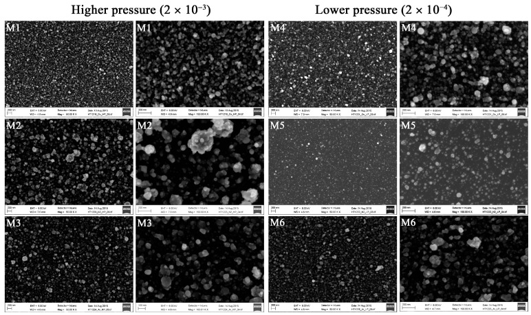
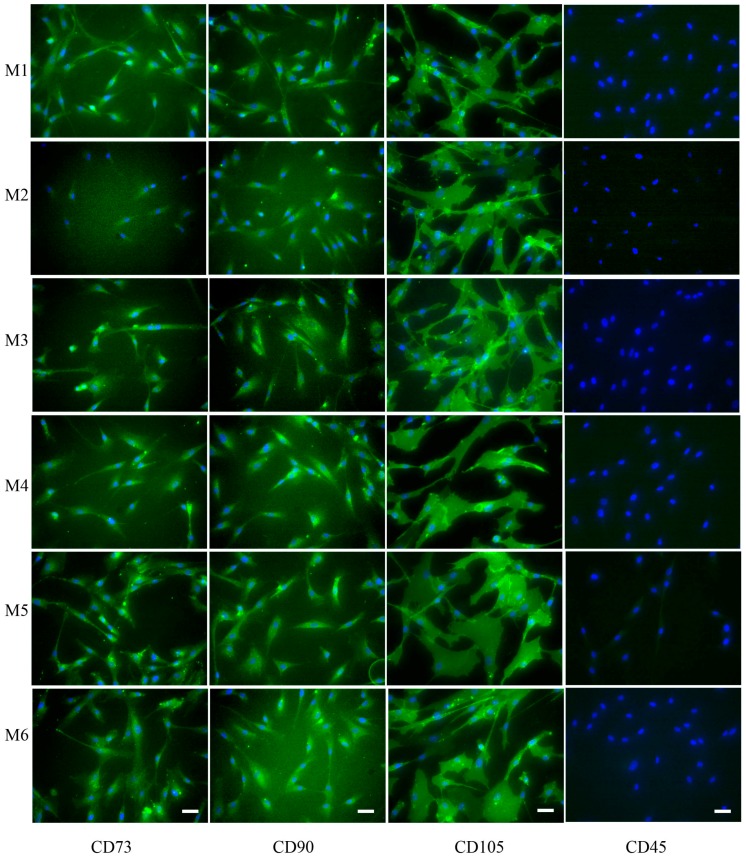
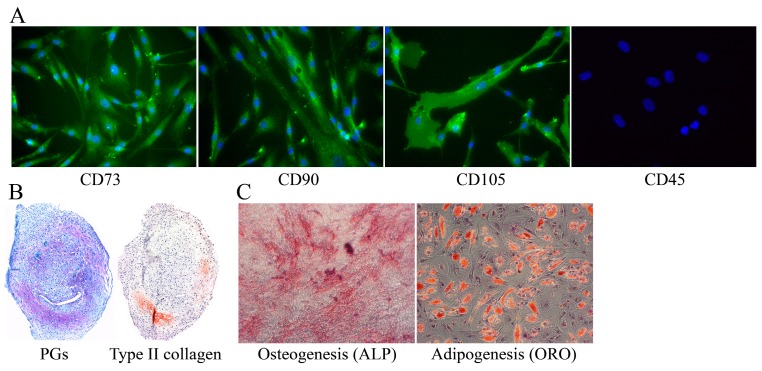
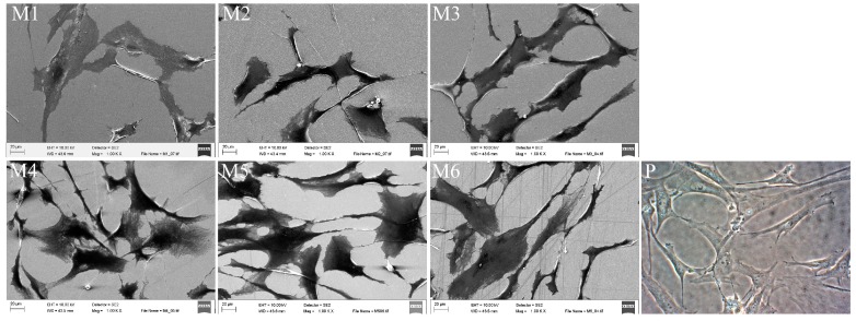
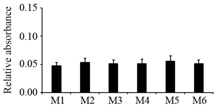
The chemical composition and texture of titanium coatings can influence the growth characteristics of the adhered cells. An enhanced proliferation of the human mesenchymal stem cells (hMSCs) would be beneficial. The present study was aimed to investigate whether titanium deposited at different atmospheres would affect the cell growth properties, cellular morphology, and expression of surface markers of hMSCs. Titanium-based coatings were deposited on silicon wafers under oxygen, nitrogen, or argon atmospheres by ultra-short pulsed laser deposition using two different gas pressures followed by heating at 400 °C for 2 h. The characteristics of the coated surfaces were determined via contact angle, zeta potential, and scanning electron microscopy (SEM) techniques. Human MSCs were cultivated on differently coated silicon wafers for 48 h. Subsequently, the cell proliferation rates were analyzed with an MTT assay. The phenotype of hMSCs was checked via immunocytochemical stainings of MSC-associated markers CD73, CD90, and CD105, and the adhesion, spreading, and morphology of hMSCs on coated materials via SEM. The cell proliferation rates of the hMSCs were similar on all coated silicon wafers. The hMSCs retained the MSC phenotype by expressing MSC-associated markers and fibroblast-like morphology with cellular projections. Furthermore, no significant differences could be found in the size of the cells when cultured on all various coated surfaces. In conclusion, despite certain differences in the contact angles and the zeta potentials of various titanium-based coatings, no single coating markedly improved the growth characteristics of hMSCs.
钛涂层的化学成分和质地会影响黏附细胞的生长特性。人骨髓间充质干细胞(hMSCs)增殖的增强将是有益的。本研究旨在调查在不同气氛下沉积的钛是否会影响hMSCs的细胞生长特性、细胞形态以及表面标志物的表达。通过超短脉冲激光沉积,在氧气、氮气或氩气气氛下,使用两种不同的气压,在硅片上沉积钛基涂层,然后在400°C下加热2小时。通过接触角、zeta电位和扫描电子显微镜(SEM)技术确定涂层表面的特性。将人骨髓间充质干细胞在不同涂层的硅片上培养48小时。随后,用MTT法分析细胞增殖率。通过对骨髓间充质干细胞相关标志物CD73、CD90和CD105进行免疫细胞化学染色来检查hMSCs的表型,并通过扫描电子显微镜观察hMSCs在涂层材料上的黏附、铺展和形态。在所有涂层硅片上,hMSCs的细胞增殖率相似。hMSCs通过表达骨髓间充质干细胞相关标志物和具有细胞突起的成纤维细胞样形态而保留了骨髓间充质干细胞的表型。此外,在所有不同涂层表面上培养时,细胞大小没有发现显著差异。总之,尽管各种钛基涂层的接触角和zeta电位存在一定差异,但没有一种涂层能显著改善hMSCs的生长特性。