Chen J-Y, Luo C-W, Lai Y-S, Wu C-C, Hung W-C
National Institute of Cancer Research, National Health Research Institutes, Tainan, Taiwan.
Department of Pathology, Kaohsiung Medical University Hospital, Kaohsiung Medical University, Kaohsiung, Taiwan.
Oncogenesis. 2017 Aug 7;6(8):e369. doi: 10.1038/oncsis.2017.71.
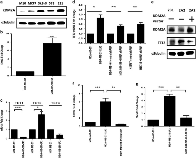
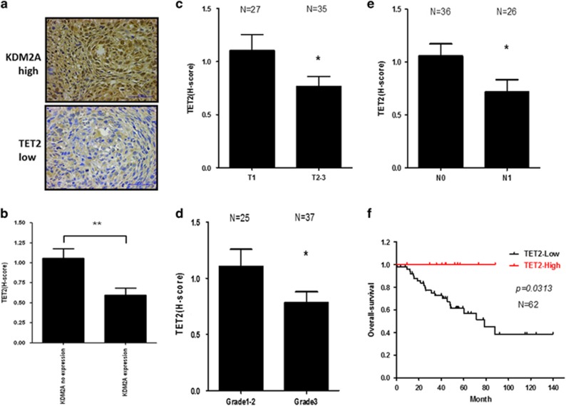
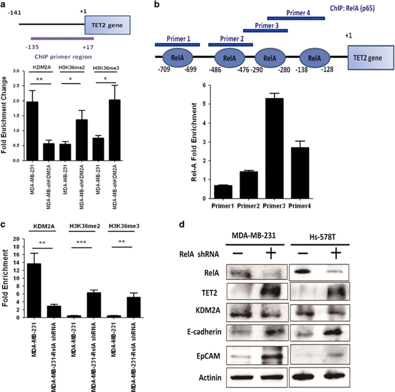
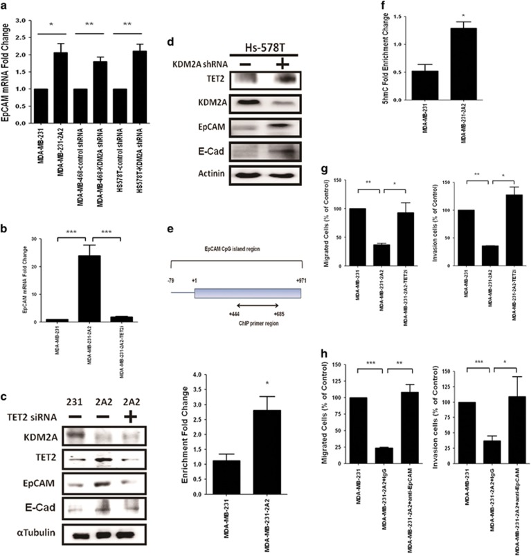
The coupling between DNA methylation and histone modification contributes to aberrant expression of oncogenes or tumor suppressor genes that leads to tumor development. Our previous study demonstrated that lysine demethylase 2A (KDM2A) functions as an oncogene in breast cancer by promoting cancer stemness and angiogenesis via activation of the Notch signaling. Here, we demonstrate that knockdown of KDM2A significantly increases the 5'-hydroxymethylcytosine (5'-hmc) level in genomic DNA and expression of tet-eleven translocation 2 (TET2) in various breast cancer cell lines. Conversely, ectopic expression of KDM2A inhibits TET2 expression in KDM2A-depleted cells suggesting TET2 is a transcriptional repression target of KDM2A. Our results show that KDM2A interacts with RelA to co-occupy at the TET2 gene promoter to repress transcription and depletion of RelA or KDM2A restores TET2 expression. Upregulation of TET2 in the KDM2A-depleted cells induces the re-activation of two TET downstream tumor suppressor genes, epithelial cell adhesion molecule (EpCAM) and E-cadherin, and inhibits migration and invasion. On the contrary, knockdown of TET2 in these cells decreases EpCAM and E-cadherin and increases cell invasiveness. More importantly, TET2 expression is negatively associated KDM2A in triple-negative breast tumor tissues, and its expression predicts a better survival. Taken together, we demonstrate for the first time that TET2 is a direct repression target of KDM2A and reveal a novel mechanism by which KDM2A promotes DNA methylation and breast cancer progression via the inhibition of a DNA demethylase.
DNA甲基化与组蛋白修饰之间的偶联作用会导致癌基因或肿瘤抑制基因的异常表达,进而引发肿瘤发展。我们之前的研究表明,赖氨酸去甲基化酶2A(KDM2A)在乳腺癌中作为一种癌基因发挥作用,它通过激活Notch信号通路促进癌症干性和血管生成。在此,我们证明,敲低KDM2A可显著提高多种乳腺癌细胞系基因组DNA中的5'-羟甲基胞嘧啶(5'-hmc)水平以及tet-eleven易位蛋白2(TET2)的表达。相反,KDM2A的异位表达会抑制KDM2A缺失细胞中TET2的表达,这表明TET2是KDM2A的转录抑制靶点。我们的结果显示,KDM2A与RelA相互作用,共同占据TET2基因启动子以抑制转录,而RelA或KDM2A的缺失可恢复TET2的表达。KDM2A缺失细胞中TET2的上调会诱导两个TET下游肿瘤抑制基因上皮细胞粘附分子(EpCAM)和E-钙粘蛋白的重新激活,并抑制迁移和侵袭。相反,在这些细胞中敲低TET2会降低EpCAM和E-钙粘蛋白的水平并增加细胞侵袭性。更重要的是,在三阴性乳腺肿瘤组织中,TET2的表达与KDM2A呈负相关,并且其表达预示着更好的生存率。综上所述,我们首次证明TET2是KDM2A的直接抑制靶点,并揭示了一种新的机制,即KDM2A通过抑制DNA去甲基化酶促进DNA甲基化和乳腺癌进展。