Boddaert N, Salvador A, Chandesris M O, Lemaître H, Grévent D, Gauthier C, Naggara O, Georgin-Lavialle S, Moura D S, Munsch F, Jaafari N, Zilbovicius M, Lortholary O, Gaillard R, Hermine O
Department of Pediatric Radiology, Hôpital Necker-Enfants Malades, AP-HP, University René Descartes, PRES Sorbonne Paris Cité, INSERM U1000 and UMR 1163, Institut Imagine, Paris, France.
Laboratoire de "Physiopathologie des Maladies Psychiatriques", Centre de Psychiatrie et Neurosciences U894, INSERM, Université Paris Descartes, Sorbonne Paris Cité, Paris, France.
Transl Psychiatry. 2017 Aug 8;7(8):e1197. doi: 10.1038/tp.2017.137.
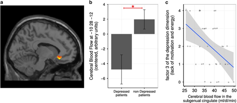
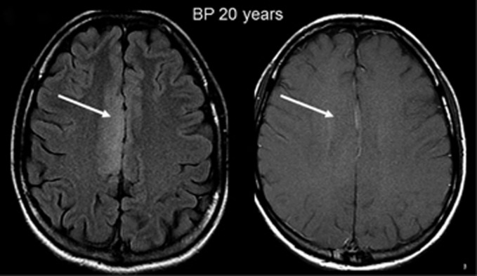
Mastocytosis is a rare disease in which chronic symptoms are related to mast cell accumulation and activation. Patients can display depression-anxiety-like symptoms and cognitive impairment. The pathophysiology of these symptoms may be associated with tissular mast cell infiltration, mast cell mediator release or both. The objective of this study is to perform morphological or functional brain analyses in mastocytosis to identify brain changes associated with this mast cell disorder. We performed a prospective and monocentric comparative study to evaluate the link between subjective psycho-cognitive complaints, psychiatric evaluation and objective medical data using magnetic resonance imaging with morphological and perfusion sequences (arterial spin-labeled perfusion) in 39 patients with mastocytosis compared with 33 healthy controls. In the test cohort of 39 mastocytosis patients with psycho-cognitive complaints, we found that 49% of them had morphological brain abnormalities, mainly abnormal punctuated white matter abnormalities (WMA). WMA were equally frequent in cutaneous mastocytosis patients and indolent forms of systemic mastocytosis patients (42% and 41% of patients with WMA, respectively). Patients with WMA showed increased perfusion in the putamen compared with patients without WMA and with healthy controls. Putamen perfusion was also negatively correlated with depression subscores. This study demonstrates, for we believe the first time, a high prevalence of morphological and functional abnormalities in the brains of mastocytosis patients with neuropsychiatric complaints. Further studies are required to determine the mechanism underpinning this association and to ascertain its specificity.
肥大细胞增多症是一种罕见疾病,其慢性症状与肥大细胞的积累和激活有关。患者可能会出现类似抑郁焦虑的症状和认知障碍。这些症状的病理生理学可能与组织中肥大细胞浸润、肥大细胞介质释放或两者都有关。本研究的目的是对肥大细胞增多症患者进行脑部形态学或功能分析,以确定与这种肥大细胞疾病相关的脑部变化。我们进行了一项前瞻性单中心比较研究,使用磁共振成像的形态学和灌注序列(动脉自旋标记灌注),评估39例肥大细胞增多症患者与33名健康对照者的主观心理认知主诉、精神科评估和客观医学数据之间的联系。在39例有心理认知主诉的肥大细胞增多症患者的测试队列中,我们发现其中49%有脑部形态学异常,主要是点状白质异常(WMA)。皮肤肥大细胞增多症患者和惰性系统性肥大细胞增多症患者的WMA出现频率相同(分别为42%和41%的患者有WMA)。与没有WMA的患者和健康对照相比,有WMA的患者壳核灌注增加。壳核灌注也与抑郁亚评分呈负相关。我们认为,这项研究首次证明了有神经精神主诉的肥大细胞增多症患者脑部形态学和功能异常的高患病率。需要进一步研究来确定这种关联的潜在机制并确定其特异性。