• 文献检索
  • 文档翻译
  • 深度研究
  • 学术资讯
  • Suppr Zotero 插件Zotero 插件
  • 邀请有礼
  • 套餐&价格
  • 历史记录
应用&插件
Suppr Zotero 插件Zotero 插件浏览器插件Mac 客户端Windows 客户端微信小程序
定价
高级版会员购买积分包购买API积分包
服务
文献检索文档翻译深度研究API 文档MCP 服务
关于我们
关于 Suppr公司介绍联系我们用户协议隐私条款
关注我们

Suppr 超能文献

核心技术专利:CN118964589B侵权必究
粤ICP备2023148730 号-1Suppr @ 2026

文献检索

告别复杂PubMed语法,用中文像聊天一样搜索,搜遍4000万医学文献。AI智能推荐,让科研检索更轻松。

立即免费搜索

文件翻译

保留排版,准确专业,支持PDF/Word/PPT等文件格式,支持 12+语言互译。

免费翻译文档

深度研究

AI帮你快速写综述,25分钟生成高质量综述,智能提取关键信息,辅助科研写作。

立即免费体验

三个 TF 共表达模块调节雄性小鼠压力超负荷性心肌肥厚。

Three TF Co-expression Modules Regulate Pressure-Overload Cardiac Hypertrophy in Male Mice.

机构信息

Biodiversity Research Center, Academia Sinica, Taipei, Taiwan.

Institute of Biomedical Sciences, Academia Sinica, Taipei, Taiwan.

出版信息

Sci Rep. 2017 Aug 8;7(1):7560. doi: 10.1038/s41598-017-07981-4.

DOI:10.1038/s41598-017-07981-4
PMID:28790436
原文链接:https://pmc.ncbi.nlm.nih.gov/articles/PMC5548763/
Abstract

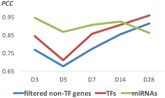
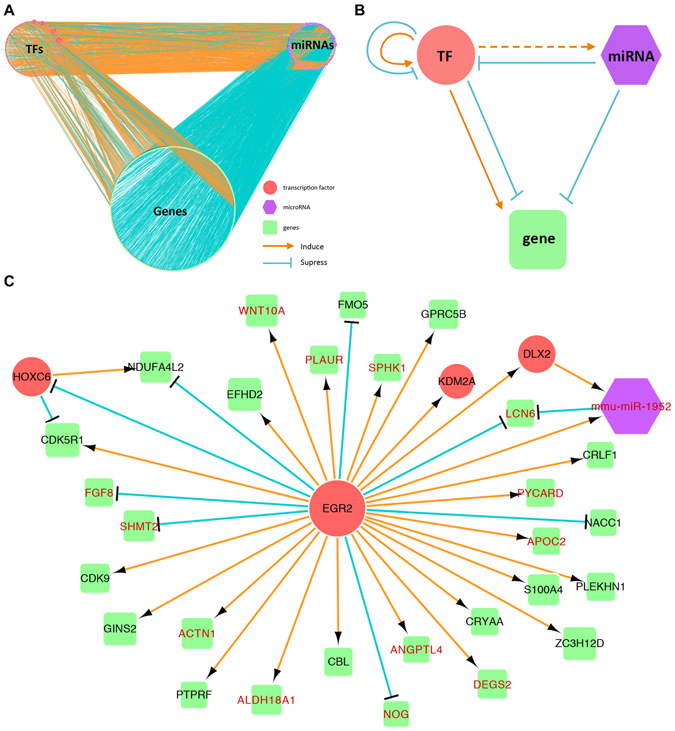
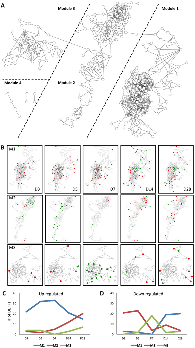
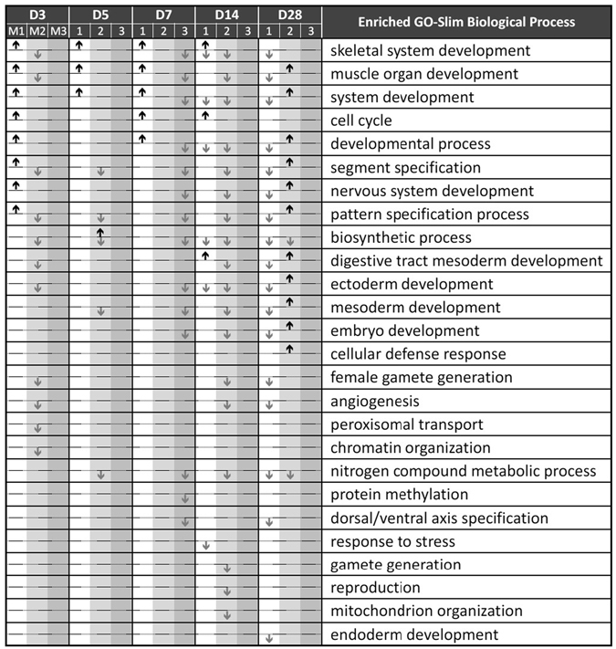
Pathological cardiac hypertrophy, a dynamic remodeling process, is a major risk factor for heart failure. Although a number of key regulators and related genes have been identified, how the transcription factors (TFs) dynamically regulate the associated genes and control the morphological and electrophysiological changes during the hypertrophic process are still largely unknown. In this study, we obtained the time-course transcriptomes at five time points in four weeks from male murine hearts subjected to transverse aorta banding surgery. From a series of computational analyses, we identified three major co-expression modules of TF genes that may regulate the gene expression changes during the development of cardiac hypertrophy in mice. After pressure overload, the TF genes in Module 1 were up-regulated before the occurrence of significant morphological changes and one week later were down-regulated gradually, while those in Modules 2 and 3 took over the regulation as the heart size increased. Our analyses revealed that the TF genes up-regulated at the early stages likely initiated the cascading regulation and most of the well-known cardiac miRNAs were up-regulated at later stages for suppression. In addition, the constructed time-dependent regulatory network reveals some TFs including Egr2 as new candidate key regulators of cardiovascular-associated (CV) genes.

摘要

病理性心肌肥厚是一种动态重塑过程,是心力衰竭的主要危险因素。尽管已经确定了许多关键调节因子和相关基因,但转录因子(TFs)如何动态调节相关基因,并在肥厚过程中控制形态和电生理变化,在很大程度上仍然未知。在这项研究中,我们从接受横主动脉结扎手术的雄性小鼠心脏中获得了四个星期内五个时间点的时间过程转录组。通过一系列计算分析,我们鉴定出三个主要的 TF 基因共表达模块,这些模块可能调节小鼠心肌肥厚发育过程中的基因表达变化。在压力超负荷后,模块 1 中的 TF 基因在发生显著形态变化之前上调,并在一周后逐渐下调,而模块 2 和 3 中的 TF 基因则在心脏增大时接管调节。我们的分析表明,早期上调的 TF 基因可能启动了级联调节,而大多数已知的心脏 miRNA 在后期被上调以进行抑制。此外,构建的时变调控网络揭示了一些 TFs,包括 Egr2,作为心血管相关(CV)基因的新候选关键调控因子。

https://cdn.ncbi.nlm.nih.gov/pmc/blobs/d447/5548763/3d27a3ad4ab4/41598_2017_7981_Fig5_HTML.jpg
https://cdn.ncbi.nlm.nih.gov/pmc/blobs/d447/5548763/0cc91e43fe24/41598_2017_7981_Fig1_HTML.jpg
https://cdn.ncbi.nlm.nih.gov/pmc/blobs/d447/5548763/bce309fcfcac/41598_2017_7981_Fig2_HTML.jpg
https://cdn.ncbi.nlm.nih.gov/pmc/blobs/d447/5548763/c5b64e1553ec/41598_2017_7981_Fig3_HTML.jpg
https://cdn.ncbi.nlm.nih.gov/pmc/blobs/d447/5548763/1f3abfa9cc20/41598_2017_7981_Fig4_HTML.jpg
https://cdn.ncbi.nlm.nih.gov/pmc/blobs/d447/5548763/3d27a3ad4ab4/41598_2017_7981_Fig5_HTML.jpg

相似文献

1
Three TF Co-expression Modules Regulate Pressure-Overload Cardiac Hypertrophy in Male Mice.三个 TF 共表达模块调节雄性小鼠压力超负荷性心肌肥厚。
Sci Rep. 2017 Aug 8;7(1):7560. doi: 10.1038/s41598-017-07981-4.
2
Comparative analysis of mRNA isoform expression in cardiac hypertrophy and development reveals multiple post-transcriptional regulatory modules.比较分析心肌肥厚和发育过程中 mRNA 异构体的表达,揭示了多个转录后调控模块。
PLoS One. 2011;6(7):e22391. doi: 10.1371/journal.pone.0022391. Epub 2011 Jul 22.
3
Systems biology approach identifies key regulators and the interplay between miRNAs and transcription factors for pathological cardiac hypertrophy.系统生物学方法鉴定病理性心肌肥厚的关键调控因子及 miRNA 和转录因子之间的相互作用。
Gene. 2019 May 25;698:157-169. doi: 10.1016/j.gene.2019.02.056. Epub 2019 Mar 5.
4
The histone demethylase PHF8 represses cardiac hypertrophy upon pressure overload.组蛋白去甲基化酶PHF8在压力超负荷时抑制心肌肥大。
Exp Cell Res. 2015 Jul 1;335(1):123-34. doi: 10.1016/j.yexcr.2015.04.012. Epub 2015 Apr 25.
5
Disruption of actin dynamics regulated by Rho effector mDia1 attenuates pressure overload-induced cardiac hypertrophic responses and exacerbates dysfunction.由Rho效应器mDia1调节的肌动蛋白动力学破坏减弱了压力超负荷诱导的心脏肥厚反应并加剧了功能障碍。
Cardiovasc Res. 2021 Mar 21;117(4):1103-1117. doi: 10.1093/cvr/cvaa206.
6
Gene expression profiling of hypertrophic cardiomyocytes identifies new players in pathological remodelling.肥厚型心肌病细胞基因表达谱分析揭示病理重构中的新角色。
Cardiovasc Res. 2021 May 25;117(6):1532-1545. doi: 10.1093/cvr/cvaa233.
7
Effects of pressure overload on extracellular matrix expression in the heart of the atrial natriuretic peptide-null mouse.压力超负荷对心钠素基因敲除小鼠心脏细胞外基质表达的影响。
Hypertension. 2003 Jul;42(1):88-95. doi: 10.1161/01.HYP.0000074905.22908.A6. Epub 2003 May 19.
8
Augmented cardiac hypertrophy in response to pressure overload in mice lacking the prostaglandin I2 receptor.在缺乏前列腺素I2受体的小鼠中,对压力超负荷的反应增强的心脏肥大。
Circulation. 2005 Jul 5;112(1):84-92. doi: 10.1161/CIRCULATIONAHA.104.527077. Epub 2005 Jun 27.
9
Mixed lineage kinase-3 prevents cardiac dysfunction and structural remodeling with pressure overload.混合谱系激酶-3 可预防心脏压力超负荷导致的心脏功能障碍和结构重塑。
Am J Physiol Heart Circ Physiol. 2019 Jan 1;316(1):H145-H159. doi: 10.1152/ajpheart.00029.2018. Epub 2018 Oct 26.
10
Induction of heart failure by minimally invasive aortic constriction in mice: reduced peroxisome proliferator-activated receptor γ coactivator levels and mitochondrial dysfunction.经微创主动脉缩窄诱导的小鼠心力衰竭:过氧化物酶体增殖物激活受体 γ 共激活因子水平降低和线粒体功能障碍。
J Thorac Cardiovasc Surg. 2011 Feb;141(2):492-500, 500.e1. doi: 10.1016/j.jtcvs.2010.03.029. Epub 2010 May 5.

引用本文的文献

1
Sirtuin 4 accelerates heart failure development by enhancing reactive oxygen species-mediated profibrotic transcriptional signaling.沉默调节蛋白4通过增强活性氧介导的促纤维化转录信号来加速心力衰竭的发展。
J Mol Cell Cardiol Plus. 2025 May 6;12:100299. doi: 10.1016/j.jmccpl.2025.100299. eCollection 2025 Jun.
2
The microRNA-204-5p inhibits APJ signalling and confers resistance to cardiac hypertrophy and dysfunction.miR-204-5p 抑制 APJ 信号转导,从而导致心脏肥厚和功能障碍的抗性。
Clin Transl Med. 2022 Jan;12(1):e693. doi: 10.1002/ctm2.693.
3
Transcriptional circuitry atlas of genetic diverse unstimulated murine and human macrophages define disparity in population-wide innate immunity.

本文引用的文献

1
Runx1 Deficiency Protects Against Adverse Cardiac Remodeling After Myocardial Infarction.Runx1 缺乏可防止心肌梗死后心脏重构不良。
Circulation. 2018 Jan 2;137(1):57-70. doi: 10.1161/CIRCULATIONAHA.117.028911. Epub 2017 Oct 13.
2
Cardiac nonmyocytes in the hub of cardiac hypertrophy.心脏肥大中心的心脏非心肌细胞。
Circ Res. 2015 Jun 19;117(1):89-98. doi: 10.1161/CIRCRESAHA.117.305349.
3
Perivascular Gli1+ progenitors are key contributors to injury-induced organ fibrosis.血管周围Gli1+祖细胞是损伤诱导器官纤维化的关键促成因素。
遗传多样化未刺激的鼠源和人源巨噬细胞转录调控回路图谱定义了群体范围固有免疫的差异。
Sci Rep. 2021 Apr 1;11(1):7373. doi: 10.1038/s41598-021-86742-w.
4
Analysis of Differential Gene Expression in Three Common Rat Models of Diastolic Dysfunction.三种常见舒张功能障碍大鼠模型中的差异基因表达分析
Front Cardiovasc Med. 2018 Feb 21;5:11. doi: 10.3389/fcvm.2018.00011. eCollection 2018.
Cell Stem Cell. 2015 Jan 8;16(1):51-66. doi: 10.1016/j.stem.2014.11.004. Epub 2014 Nov 20.
4
Identification of regulatory elements directing miR-23a-miR-27a-miR-24-2 transcriptional regulation in response to muscle hypertrophic stimuli.鉴定在响应肌肉肥大刺激时指导miR-23a-miR-27a-miR-24-2转录调控的调控元件。
Biochim Biophys Acta. 2014 Sep;1839(9):885-97. doi: 10.1016/j.bbagrm.2014.07.009. Epub 2014 Jul 19.
5
Early growth response 1 is an early signal inducing Cav3.2 T-type calcium channels during cardiac hypertrophy.早期生长反应因子 1 是在心脏肥大过程中诱导 Cav3.2 T 型钙通道的早期信号。
Cardiovasc Res. 2013 Nov 1;100(2):222-30. doi: 10.1093/cvr/cvt190. Epub 2013 Aug 8.
6
Mending broken hearts: cardiac development as a basis for adult heart regeneration and repair.修复破损的心脏:心脏发育为成年心脏再生和修复的基础。
Nat Rev Mol Cell Biol. 2013 Aug;14(8):529-41. doi: 10.1038/nrm3619. Epub 2013 Jul 10.
7
MicroRNA-26 was decreased in rat cardiac hypertrophy model and may be a promising therapeutic target.微小 RNA-26 在大鼠心肌肥厚模型中表达降低,可能是一个有前途的治疗靶点。
J Cardiovasc Pharmacol. 2013 Sep;62(3):312-9. doi: 10.1097/FJC.0b013e31829b82e6.
8
Architecture of the human regulatory network derived from ENCODE data.人类调控网络的结构源自 ENCODE 数据。
Nature. 2012 Sep 6;489(7414):91-100. doi: 10.1038/nature11245.
9
Genetics of human cardiovascular disease.人类心血管疾病的遗传学。
Cell. 2012 Mar 16;148(6):1242-57. doi: 10.1016/j.cell.2012.03.001.
10
Differential expression of microRNAs in different disease states.microRNAs 在不同疾病状态下的差异表达。
Circ Res. 2012 Feb 17;110(4):638-50. doi: 10.1161/CIRCRESAHA.111.247437.