• 文献检索
  • 文档翻译
  • 深度研究
  • 学术资讯
  • Suppr Zotero 插件Zotero 插件
  • 邀请有礼
  • 套餐&价格
  • 历史记录
应用&插件
Suppr Zotero 插件Zotero 插件浏览器插件Mac 客户端Windows 客户端微信小程序
定价
高级版会员购买积分包购买API积分包
服务
文献检索文档翻译深度研究API 文档MCP 服务
关于我们
关于 Suppr公司介绍联系我们用户协议隐私条款
关注我们

Suppr 超能文献

核心技术专利:CN118964589B侵权必究
粤ICP备2023148730 号-1Suppr @ 2026

文献检索

告别复杂PubMed语法,用中文像聊天一样搜索,搜遍4000万医学文献。AI智能推荐,让科研检索更轻松。

立即免费搜索

文件翻译

保留排版,准确专业,支持PDF/Word/PPT等文件格式,支持 12+语言互译。

免费翻译文档

深度研究

AI帮你快速写综述,25分钟生成高质量综述,智能提取关键信息,辅助科研写作。

立即免费体验

光学相干断层扫描血管造影:青光眼诊断与研究的新工具。

Optical Coherence Tomography Angiography: A New Tool in Glaucoma Diagnostics and Research.

作者信息

Daneshvar Ramin, Nouri-Mahdavi Kouros

机构信息

Glaucoma Division, Stein Eye Institute, David Geffen School of Medicine, University of California Los Angeles, Los Angeles, California, USA.

Eye Research Center, Mashhad University of Medical Sciences, Mashhad, Iran.

出版信息

J Ophthalmic Vis Res. 2017 Jul-Sep;12(3):325-332. doi: 10.4103/jovr.jovr_36_17.

DOI:10.4103/jovr.jovr_36_17
PMID:28791067
原文链接:https://pmc.ncbi.nlm.nih.gov/articles/PMC5525503/
Abstract

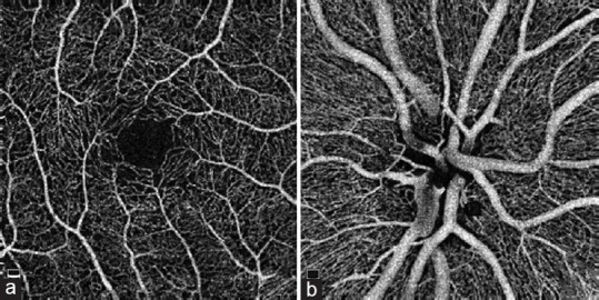
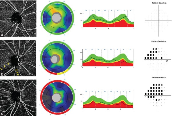
Optical coherence tomography angiography (OCTA) is a new modality in ocular imaging which provides high resolution view of the vascular structures in the retina and optic nerve head. This technology has the advantages of being noninvasive, rapid and reproducible. OCTA is becoming a valuable tool for evaluating many retinal and optic nerve diseases. This article provides a brief introduction to the technology and its application in the field of glaucoma diagnostics.

摘要

光学相干断层扫描血管造影(OCTA)是眼部成像的一种新方式,它能提供视网膜和视神经乳头血管结构的高分辨率视图。这项技术具有非侵入性、快速且可重复的优点。OCTA正成为评估多种视网膜和视神经疾病的重要工具。本文简要介绍了该技术及其在青光眼诊断领域的应用。

https://cdn.ncbi.nlm.nih.gov/pmc/blobs/8711/5525503/df01936dfc6a/JOVR-12-325-g003.jpg
https://cdn.ncbi.nlm.nih.gov/pmc/blobs/8711/5525503/b6c9a8fbbd17/JOVR-12-325-g001.jpg
https://cdn.ncbi.nlm.nih.gov/pmc/blobs/8711/5525503/df01936dfc6a/JOVR-12-325-g003.jpg
https://cdn.ncbi.nlm.nih.gov/pmc/blobs/8711/5525503/b6c9a8fbbd17/JOVR-12-325-g001.jpg
https://cdn.ncbi.nlm.nih.gov/pmc/blobs/8711/5525503/df01936dfc6a/JOVR-12-325-g003.jpg

相似文献

1
Optical Coherence Tomography Angiography: A New Tool in Glaucoma Diagnostics and Research.光学相干断层扫描血管造影:青光眼诊断与研究的新工具。
J Ophthalmic Vis Res. 2017 Jul-Sep;12(3):325-332. doi: 10.4103/jovr.jovr_36_17.
2
Optical Coherence Tomography Angiography in Glaucoma Care.光学相干断层扫描血管造影在青光眼治疗中的应用。
Curr Eye Res. 2018 Sep;43(9):1067-1082. doi: 10.1080/02713683.2018.1475013. Epub 2018 May 23.
3
Optical coherence tomography angiography: an overview of the technology and an assessment of applications for clinical research.光学相干断层扫描血管造影术:技术概述及临床研究应用评估
Br J Ophthalmol. 2017 Jan;101(1):16-20. doi: 10.1136/bjophthalmol-2016-309389. Epub 2016 Oct 4.
4
Potential applications of optical coherence tomography angiography in glaucoma.光学相干断层扫描血管造影术在青光眼方面的潜在应用
Curr Opin Ophthalmol. 2018 May;29(3):226-233. doi: 10.1097/ICU.0000000000000475.
5
[Optical coherence tomography angiography: Value for glaucoma diagnostics].[光学相干断层扫描血管造影术:在青光眼诊断中的价值]
Ophthalmologe. 2019 Jul;116(7):602-609. doi: 10.1007/s00347-018-0815-9.
6
Optical Coherence Tomography Angiography to Better understand Glaucoma.光学相干断层扫描血管造影术以更好地了解青光眼。
J Curr Glaucoma Pract. 2017 May-Aug;11(2):35-37. doi: 10.5005/jp-journals-10028-1219. Epub 2017 Aug 5.
7
A Review of OCT Angiography in Glaucoma.青光眼的光学相干断层扫描血管造影综述
Semin Ophthalmol. 2019;34(4):279-286. doi: 10.1080/08820538.2019.1620807. Epub 2019 Jun 3.
8
Optical Coherence Tomography Angiography of the Optic Disc; an Overview.视盘光学相干断层扫描血管造影术概述
J Ophthalmic Vis Res. 2017 Jan-Mar;12(1):98-105. doi: 10.4103/2008-322X.200162.
9
Optical coherence tomography angiography in glaucoma.光学相干断层扫描血管造影术在青光眼中的应用。
Curr Opin Ophthalmol. 2019 Mar;30(2):110-116. doi: 10.1097/ICU.0000000000000554.
10
Optical Coherence Tomography Angiography and Glaucoma: A Brief Review.光学相干断层扫描血管造影与青光眼:简要综述
Asia Pac J Ophthalmol (Phila). 2019;8(2):115-125. doi: 10.22608/APO.201914.

引用本文的文献

1
Optic Nerve Head Pulsatile Displacement in Open-Angle Glaucoma after Intraocular Pressure Reduction Measured by Optical Coherence Tomography: A Pilot Study.通过光学相干断层扫描测量眼压降低后开角型青光眼中的视神经乳头搏动性移位:一项初步研究。
Bioengineering (Basel). 2024 Apr 23;11(5):411. doi: 10.3390/bioengineering11050411.
2
Mapping Pulsatile Optic Nerve Head Deformation Using OCT.使用光学相干断层扫描(OCT)绘制搏动性视神经乳头变形图。
Ophthalmol Sci. 2022 Aug 6;2(4):100205. doi: 10.1016/j.xops.2022.100205. eCollection 2022 Dec.
3
Subretinal Fibrosis Detection Using Polarization Sensitive Optical Coherence Tomography.

本文引用的文献

1
Alterations of the Foveal Avascular Zone Measured by Optical Coherence Tomography Angiography in Glaucoma Patients With Central Visual Field Defects.通过光学相干断层扫描血管造影术测量的青光眼中央视野缺损患者黄斑无血管区的改变
Invest Ophthalmol Vis Sci. 2017 Mar 1;58(3):1637-1645. doi: 10.1167/iovs.16-21079.
2
Multifaceted Presentation of Recurrent Spontaneous Coronary Artery Dissection: Angiography and Optical Coherence Tomography Findings.复发性自发性冠状动脉夹层的多方面表现:血管造影和光学相干断层扫描结果
Circ Cardiovasc Interv. 2017 Feb;10(2):e004696. doi: 10.1161/CIRCINTERVENTIONS.116.004696.
3
OPTICAL COHERENCE TOMOGRAPHY ANGIOGRAPHY OF THE FOVEA IN CHILDREN BORN PRETERM.
使用偏振敏感光学相干断层扫描检测视网膜下纤维化
Transl Vis Sci Technol. 2020 Mar 16;9(4):13. doi: 10.1167/tvst.9.4.13. eCollection 2020 Mar.
4
Parafoveal vessel changes in primary open-angle glaucoma and normal-tension glaucoma using optical coherence tomography angiography.使用光学相干断层扫描血管造影术观察原发性开角型青光眼和正常眼压性青光眼的黄斑旁血管变化
Clin Ophthalmol. 2019 Sep 27;13:1935-1945. doi: 10.2147/OPTH.S206288. eCollection 2019.
5
An overview of optical coherence tomography angiography and the posterior pole.光学相干断层扫描血管造影术与后极部概述。
Ther Adv Ophthalmol. 2019 Apr 3;11:2515841419840249. doi: 10.1177/2515841419840249. eCollection 2019 Jan-Dec.
早产儿黄斑区的光学相干断层扫描血管造影
Retina. 2017 Dec;37(12):2289-2294. doi: 10.1097/IAE.0000000000001471.
4
CLINICAL TRIAL ENDPOINTS FOR OPTICAL COHERENCE TOMOGRAPHY ANGIOGRAPHY IN NEOVASCULAR AGE-RELATED MACULAR DEGENERATION.光学相干断层扫描血管造影术用于新生血管性年龄相关性黄斑变性的临床试验终点
Retina. 2016 Dec;36 Suppl 1:S83-S92. doi: 10.1097/IAE.0000000000001338.
5
Acoustic-actuated optical coherence angiography.声学驱动光学相干血管造影术。
Opt Lett. 2016 Dec 15;41(24):5813-5816. doi: 10.1364/OL.41.005813.
6
Diagnostic ability of peripapillary vessel density measurements of optical coherence tomography angiography in primary open-angle and angle-closure glaucoma.光学相干断层扫描血管造影术测量视乳头周围血管密度在原发性开角型青光眼和闭角型青光眼中的诊断能力
Br J Ophthalmol. 2017 Aug;101(8):1066-1070. doi: 10.1136/bjophthalmol-2016-309377. Epub 2016 Nov 29.
7
Microvascular Compromise Develops Following Nerve Fiber Layer Damage in Normal-Tension Glaucoma Without Choroidal Vasculature Involvement.在无脉络膜血管受累的正常眼压青光眼中,神经纤维层损伤后会发生微血管损害。
J Glaucoma. 2017 Mar;26(3):216-222. doi: 10.1097/IJG.0000000000000587.
8
Comparison of coronary arterial lumen dimensions on angiography and plaque characteristics on optical coherence tomography images and their changes induced by statin.血管造影术测定的冠状动脉管腔尺寸与光学相干断层扫描图像上的斑块特征及其他汀类药物诱导变化的比较。
BMC Med Imaging. 2016 Nov 22;16(1):63. doi: 10.1186/s12880-016-0166-4.
9
OCT Angiography of the Peripapillary Retina in Primary Open-Angle Glaucoma.原发性开角型青光眼视乳头周围视网膜的光学相干断层扫描血管造影
Invest Ophthalmol Vis Sci. 2016 Nov 1;57(14):6265-6270. doi: 10.1167/iovs.16-20287.
10
A review of optical coherence tomography angiography (OCTA).光学相干断层扫描血管造影术(OCTA)综述。
Int J Retina Vitreous. 2015 Apr 15;1:5. doi: 10.1186/s40942-015-0005-8. eCollection 2015.